3.7.5...6 3 Metabolism 138
of a free radical at the deoxyadenosyl group. This radical is used 3.7.6 Folate and Pterines
to exchange the positions of hydrogen and various substituents One vitamin and several cofactors contain a pteridine ring system:
at neighboring atoms.
• Folate/tetrahydrofolate/tetrahydrofolylpolyglutamate (THF)
Examples: methylmalonyl-CoA mutase in mammals • Biopterin/tetrahydrobiopterin (THB)
(Fig. 3.2.5-2, exchanging H and a - CO-S-CoA group), propio- • Molybdenum (and tungsten) cofactors (MoCo)
nate and glutamate fermentation by bacteria (Figs. 3.10.5-2 and • Methanopterin/tetrahydromethanopterin (THMPT).
3.10.5-4).
- Reduction of ribonucleotides: Some bacterial ribonucleoside- The synthesis of folate compounds is restricted to plants and micro-
triphosphate reductases (3.6.1-4) contain coenzyme B12 instead organisms, but the product is required as a vitamin (vitamin Bc) by
of the Fe(III)-O2−-Fe(III) center to generate a free radical. animals. Biopterin synthesis seems to be only performed by animals,
whereas the molybdenum cofactor can apparently be synthesized by
• Heterolytic cleavage, employing methyl cobalamin: In some all living organisms. Methanopterin is formed exclusively by anaero-
bic archaea (methanogens).
methyl transferases, methylcobalamin acts as the methyl group
donor (Co3+-CH3 Æ Co+ + CH3, Fig. 3.7.5-3). 3.7.6.1 Tetrahydrofolate/Folylpolyglutamate (Fig. 3.7.6-1)
Biosynthesis: The synthesis of folate starts with a two step conversion of
Examples are homocysteine methyltransferases in mammals GTP to dihydroneopterin-P3 by GTP cyclohydrolase I (GTPCH I). C-8 of
and bacteria (Fig. 3.2.5-2). B12 is then remethylated by 5-methyl- GTP is released as formate. The enzyme, in contrast to GTPCH II (ribo-
tetrahydrofolate. An occasional oxidation of the cofactor from flavin synthesis, 3.7.3), incorporates C atoms of ribose (1¢, 2¢) into the
the intermediate Co+ to the Co++ state requires a regeneration pteridine ring system. Dihydroneopterin-P3 is also an intermediate of the
step, involving reduction by NADPH via flavo- or ferredoxin pathways leading to biopterin (3.7.6.3) and to methanopterin (3.7.6.5).
and methylation by 5-adenosylmethionine. Related reactions are
methyl transfers in archaeal methanogenesis and bacterial aceto- The specific part of the tetrahydrofolate biosynthesis proceeds via
genesis (Figs. 3.10.6-2 and 3.10.6-3) a (so far unspecified) removal of all phosphates, side chain shortening
and pyrophosphorylation to 2-amino-4-hydroxy-6-hydroxymethyl-
3.7.5.3 Siroheme and Coenzyme F430 7,8-dihydropteridine-P2. The fusion of this intermediate with 4-ami-
Siroheme (Fig. 3.3-1) is derived from precorrin 2 by introduction of nobenzoate to 7,8-dihydropteroate is the target of the antibacterial
an iron atom. It is the coenzyme of bacterial sulfite and nitrite reduct- action of sulfonamides. After the ATP dependent addition of gluta-
ases (3.10.6). Coenzyme F430 (Fig. 3.3-1) is a nickel-containing deriv- mate, the resulting dihydrofolate is reduced to tetrahydrofolate. In
ative of precorrin 3A. It acts as cofactor in the methyl-coenzyme M eukarya, the synthesis is performed in mitochondria. Some steps are
reductase reaction of methanogenesis (3.10.6.2) and functions similar catalyzed by species specific multifunctional enzymes.
to coenzyme B12 in methyl transfer reactions.
Literature: Polyglutamylation of reduced folate: Tetrahydrofolylpolyglutamate
Banerjee, R. Chemistry and Biology 1997;4:175–186. synthase catalyzes the sequential addition of usually 2 … 8 glutamate
Blanche, F. et al. Angew. Chem. Int. Ed. 1995;34:1001–1029. residues via g-peptide linkages. Polyglutamylation increases or even
Drennan, C.L. et al. Science 1994;266:1669–1674. enables the cofactor function of folates. It also inhibits the diffusion of
Kräutler, B. Biochem. Soc. Transactions 2005;33:806–810 folates through membranes and thus enables their accumulation and
Ludwig, M.L., Matthews, R.G. Ann. Rev. of Biochem. subcellular localization. For resorption of folates from the diet, these
bonds must be hydrolyzed by pteroyl-poly-g-glutamate hydrolase.
1997;66:269–313. The dietary requirement for humans is 0.4 mg/day.
Stubbe, J. Science 1994;266:1663–1664.
Biochemical functions of folylpolyglutamate: Polyglutamylated
Figure 3.7.5-2. Mechanism of B12 Dependent Mutase Reactions tetrahydrofolate (THF) is the central cofactor of the one carbon (C1)
Employing Deoxyadenosyl Cobalamin metabolism (see below).
Figure 3.7.5-3. Reaction Mechanism of Homocysteine 3.7.6.2 General Reactions of the C1 Metabolism (Fig. 3.7.6-1)
Methyltransferase Employing Methylcobalamin C1 metabolism is a key regulatory factor of catabolic and anabolic
pathways by providing a controlled flux through the C1 pool. It encom-
passes the transfer of C1 compounds and their reduction or oxidation.
CO2 fixation and biotin dependent reactions (3.7.8.2) are not usually
discussed in this context.
THF carries C1 units at the oxidation levels of formate (as 5- or
10-formyl-THF, 5,10-methenyl- or 5-formimino-THF), formaldehyde
(as 5,10-methylene-THF) and methanol (as 5-methyl-THF). The ori-
gins of the C1 units entering at the various levels are
• 10-formyl-THF: formate
• 5-formimino-THF: degradation products of histidine (3.2.8.2) and
xanthine (3.6.1.6)
• 5,10-methylene-THF (mainly in mitochondria): serine (3.2.4.1,
main supplier of C1 units), betaine degradation products
C1 Units are provided by
• 10-formyl-THF: for purine (3.6.1.1) and N-formylmethionyl-
tRNA formation (in bacteria, archaea, mitochondria and chloro-
plasts, 4.1)
• 5,10-methylene-THF: for biosyntheses of thymidine 5¢-P (3.6.2.3),
coenzyme A (3.7.7.1) and serine (3.2.4.1, mainly in the cytosol)
• 5-methyl-THF: for methionine biosynthesis (3.2.5.4). Methionine
in the form of S-adenosylmethionine serves as the donor of all
energy driven methylation reactions.
139 3 Metabolism 3.7.6
(Fig. 3.2.7-1)
6(R)-5,6,7,8-TETRA-
HYDROBIOPTERIN
(HYDROXYLASES
e.g. 3.2.7-3)
5,6,7,8-TETRAHYDROMETHANOPTERIN
(THMPT)
3.10.6.3)
(3.4.3.5)
(3.2.5.4)
6(S)-5,6,7,8-TETRAHYDROFOLATE (THF)
(3.2.8.2)
(3.6.1.4)
(3.6.2.3)
(3.7.7.1)
(3.6.1)
(4.1.3.3)
(3.4.3-5) (3.2.4-2 etc.)
(3.2.4.2)
Figure 3.7.6-1. Biosynthesis of Pterines
3.7.6 3 Metabolism 140
The reversible interconversions of C1 compounds take place by for- Two enzymes are involved in its subsequent dehydratation and reduc-
mate-THF ligase, methenyl-THF cyclohydrolase and methylene-THF tion to THB: pterin-4a-carbinolamine dehydratase (PCD) and dihydro-
dehydrogenase (NADPH, Fig. 3.7.6-1). In animals they are combined pteridine reductase (DHPR). Enzymic recycling of THB is essential for
into a trifunctional enzyme. In mitochondria, the dehydrogenase activ- phenylalanine metabolism: (1) to ensure a continuous supply of reduced
ity is NAD-dependent and controls the metabolic flux from serine/ cofactor, and (2) to prevent accumulation of harmful metabolites pro-
glycine to formate (for protein synthesis and for cytoplasmatic needs). duced by rearrangements of THB-4a-carbinolamine.
Glycine hydroxymethyltransferase (3.2.4.2) and 5-formyl-THF cycloli-
gase interconvert 5,10-methylene-THF and 5-formyl-THF. Irreversible Biochemical functions of tetrahydrobiopterin: THB is the cofactor of
reactions are catalyzed by 10-formyl-THF dehydrogenase, which the aromatic amino acid monooxygenases (phenylalanine, tyrosine and
releases THF and by methylene-THF reductase (NADPH), which leads tryptophan monooxygenases, 3.2.7.3), of nitric oxide synthase (7.7.2)
to 5-methyl-THF. In the case of cobalamin deficiency, the 5-methyl- and of glyceryl-ether hydroxylase (e.g., PAF degradation, 3.4.3.3).
THF-homocysteine hydroxymethyltransferase (3.2.5.4) is inoperative
and a deleterious accumulation of 5-methyl-THF takes place. Since THB plays a role in the biosynthesis of serotonin (3.2.7.3)
and catecholamines (3.2.7.4), defects in THB synthesis or regenera-
Most regulation mechanisms are poorly understood. 5-Formyl- tion may lead to, e.g., neurological diseases.
THF inhibits most enzymes of the C1-THF interconversions and other
C1-THF dependent reactions. S-Adenosylmethionine inhibits the Additionally THB seems to be involved in other cofactor functions,
methylene-THF reductase. e.g., in cytokine dependent proliferation of erythroid and T cells and
in interactions with the IL-2 receptor.
3.7.6.3 Tetrahydrobiopterin (Fig. 3.7.6-1)
Biosynthesis: Tetrahydrobiopterin (THB) biosynthesis proceeds anal- Neopterin: This pteridine, together with dihydroneopterin, is secreted
ogously to folate biosynthesis (3.7.6.1) up to the dihydroneopterin-P3 from monocytes/macrophages after stimulation of THB synthesis by
level. The Zn-dependent 6-pyruvoyltetrahydropterin synthase cata- interferon-g.
lyzes an internal redox transfer and eliminates the triphosphate resi-
due. Two consecutive reduction steps are performed by the NADPH Other pteridines: There is strong evidence that in algae, fungi and
dependent sepiapterin reductase. plants, a pteridine is one of the cofactors of the blue light receptor.
The rate limiting step of THB synthesis is the initial GTP cyclohy- 3.7.6.4 Molybdenum/Tungsten Cofactors (Fig. 3.7.6-2)
drolase I reaction. Feedback inhibition by THB is mediated by the Biosynthesis: Molybdopterin (MPT) synthesis likewise seems to start
GTP cyclohydrolase I feedback regulatory protein (GFRP, formerly from GTP. Ring opening and further steps (without release of formate) lead
p35). The de novo synthesis of the cyclohydrolase is suppressed by to the labile cyclic pyranopterin monophosphate (cPMP), which is sulfur-
glucocorticoids and cell-specifically stimulated by, e.g., interferon g, ylated and rearranged by MPT synthase to MPT, forming the Mo binding
interleukin-1b or other cytokines. dithiolene structure. Incorporation of Mo (delivered by the Mo uptake and
transport system) into MPT yields the very unstable molybdenum cofactor
Regeneration of THB is an essential part of the phenylalanine (MoCo). Enzymes of the xanthine oxidase family require a final matura-
hydroxylating system. During the reactions catalyzed by aromatic tion step by addition of a terminal sulfido ligand to the molybdenum center,
amino acid hydroxylases, molecular oxygen is transferred to the cor- as catalyzed by the enzyme molybdenum cofactor sulfurase. The last step
responding amino acid and THB is oxidized to THB-4a-carbinolamine. of the synthesis is the incorporation into the target enzyme, which effects
O
N CYCLIC PYRANOPTERIN CYCLIC PYRANOPTERIN MOLYBDOPTERIN
MONOPHOSPHATE
GUANOSINE HN
5'-TRIPHOSPHATE MONOPHOSPHATE O H HO OH MOLYBDOPTERIN O SH
N SYNTHASE H2N NO SYNTHASE N
H2N N O H H SH
OOO Moa Moa Moa Moa N
P OH DE E D
HO P O P O P O O O H2N N N H O
OH OH OH PPi H2N N O H H
OO
H P
OH OH MoaE HO OH
ATP
L-CYSTEINE MoaD-GLY MOLYBDOPTERIN
ADENYLYLTRANSFERASE
CYSTEINE PPi
DESULFURASE
L-ALANINE MOLYBDOPTERIN SYNTHASE ADENYLYL-MOLYBDOPTERIN
SULFURTRANSFERASE
[CYSTEINE DESULFURASE]-S-SULFANYL-L-CYS O H SH
[MoaD]-GLY-GLY N N
CYTIDYLYL MOLYBDENUM COFACTOR H SH
X
O H S Mo X H2N N N H O
N H H
ATP H S OO
MOLYBDOPTERIN-SYNTHASE P
N NH2
PPi ADENYLTRANSFERASE HO AMP
N MoO4--
[MoaD]-GLY-GLY-AMP H2N N N O
H H H NO
AMP OOO O MOLYBDOPTERIN
P MOLYBDOTRANSFERASE
HO O P O AMP
GUANYLYL MOLYBDENUM COFACTOR OH
X PPi
MOLYBDENUM COFACTOR
O S Mo X O OH OH
N HN CTP X
H H S
N O S Mo X
N MOLYBDENUM COFACTOR N H
N CYTIDYLYLTRANSFERASE N H S
H2N N N H O H2N N PPi G T P
H H O O
OO PO N
P OH H2N N H H O
H
HO O OO
P
X = O, S, OR A SECOND MOLYBDENUM COFACTOR MOLYBDENUM COFACTOR HO OH
MoaD = SULFUR CARRIER PROTEIN GUANYLYLTRANSFERASE
OH OH
Figure 3.7.6-2. Biosynthesis of the Molybdenum Cofactor
141 3 Metabolism 3.7.6...8
the stabilization of the cofactor. In bacteria, MoCo is usually converted 2-Oxoisovalerate, the transamination product of l-valine, is meth-
into a dinucleotide before incorporation into the enzyme takes place. ylated by 5,10-methylene-THF to yield 2-dehydropantoate, which is
then reduced to (R)-pantoate. An ATP-dependent condensation with
Biochemical function: All molybdoenzymes (except nitrogenase, b-alanine (which is produced from aspartate by 1-decarboxylation,
3.2.1) contain the organometallic molybdenum cofactor. Mammalian 3.2.2.4) leads to (R)-pantothenate. Since intestinal E. coli secretes
molybdoenzymes are sulfite oxidase, xanthine oxidase (3.6.1.6), alde- considerable amounts of it, this becomes an important source of the
hyde oxidase and amidoxime reductase. Molybdopterin is degraded vitamin to the mammalian host. Also, pantothenol can be oxidized to
to urothione. Defects in molybdopterin biosynthesis in humans are pantothenate. It is present in food, but is also used as a drug.
very rare. They are lethal in early childhood. In addition to the four
molybdoenzymes occuring in mammals, plants possess assimilatory The synthesis of coenzyme A (CoA) from the vitamin pantothenate
nitrate reductases which contain a molybdenum cofactor. can be performed by all living beings and begins with its phospho-
rylation. This is the committed step (= first ambiguous step), which
In plants and bacteria, a broad range of reactions are catalyzed by is regulated via allosteric inhibition by the end product CoA. Then
molybdoenzymes. Three enzyme families were defined by functional CTP-dependent condensation with l-cysteine and decarboxylation
criteria: takes place. Adenylylation and phosphorylation complete the synthe-
sis of the coenzyme. In mammals, these two steps are carried out by
• DMSO reductase family in bacteria, e.g., DMSO reductase, nitrate a bifunctional enzyme, while in plants and bacteria separate entities
exist. The active holo-acyl carrier protein (ACP, 3.4.1.1) is obtained
reductase (dissimilatory, 3.10.6), formate dehydrogenases (one of by transfer of the 4¢-phosphopantetheine residue from CoA to a serine
them contains tungsten) residue of apo-ACP.
• Xanthine oxidase family, e.g., xanthine oxidase/dehydrogenase Dietary requirement for humans: Since pantothenate is present
in practically all biological material and is, in addition to food, sup-
(3.6.1.6, contains additionally FAD), aldehyde oxidase. plied by intestinal bacteria (see above), avitaminoses are practically
unknown. The daily requirement for adults is estimated to be in the
• Sulfite oxidase family in algae and higher plants, e.g., sulfite oxi- order of 6 … 10 mg.
dase/dehydrogenase (3.10.6), nitrate reductase (assimilatory) Degradation: CoA is cleaved by a phosphodiesterase, yielding 4¢-phos-
phopantetheine and adenosine 3¢,5¢-bisphosphate. Likewise, the cleavage
Related are tungsten enzymes, which form the majority of the alde- of holo-ACP yields 4¢-phosphopantetheine (in addition to apo-ACP).
hyde ferredoxin oxidoreductase family, e.g., aldehyde ferredoxin oxi-
doreductase. 3.7.7.2 Biochemical Function
The thioester bonds between acids and CoA or pantetheine are ener-
3.7.6.5 Methanopterin (Fig. 3.7.6-1) gy-rich bonds (DG¢0 for hydrolysis = −31.5 kJ/mol, 3.1.3.3). Their
Biosynthesis: The biosynthesis of tetrahydromethanopterin (THMPT) formation corresponds to an activation of the acid for synthetic reac-
resembles that of tetrahydrofolate and apparently branches off at the tions, such as esterification, amidation (e.g., 3.1.7), anhydride forma-
dihydroneopterin-P3 level. Then S-adenosylmethionine dependent tion (e.g., Fig. 3.10.5-4), C-C bond formation (e.g., 3.1.3.3) etc. Best
methylation at C-7 takes place. The sources of the C-9 and C-9a atoms known is acetyl-CoA, the ‘activated acetic acid’ (3.1.3.3).
have not been conclusively proven. The side chain of methanopterin is
formed by the condensation of 4-aminobenzoate (3.2.7.1) with ribose. R-COOH + ATP + CoA-SH = Acyl-CoA + AMP + PPi
The mechanism resembles the indole 3-glycerol-P synthase reaction Acyl-CoA + X = Acyl-X + CoA-SH.
(3.2.7.1). Ribosyl 5-P and 2-hydroxyglutarate (obtained by reduction
of 2-oxoglutarate, Fig. 3.1.8-2) are added afterwards. During fatty acid synthesis (3.4.1.1), the pantetheine residue of ACP
acts as a ‘movable arm’ to move the bound acid from one catalytic
Biochemical function: The C1 derivatives of tetrahydromethano- center to the next. A similar task is performed by pantetheine ‘arms’ in
pterin are analogous to those of tetrahydrofolate. They are involved in non-ribosomal polypeptide synthesis (e.g., Fig. 3.10.8-1).
methane synthesis, as described in 3.10.6.2.
Literature:
Literature:
Friedrich, W. Vitamins. De Gruyter, 1988, p. 809–835.
Auerbach, G., Nar, H. Biol. Chem. 1997;378:185–192. Jackowski, S. in Neidhardt, F.C. (Ed.). Escherichia coli and
DiMarco, A.A. et al. Ann. Rev. Biochem. 1990;59:361–370.
Hille, R., Nishino, T., Bittner, F. Coord. Chem. Rev. 2011; Salmonella. 2nd Ed. ASM Press, 1996, p. 687–694.
Kupke, T., Hernández-Acosta, P., Culiáñez-Macià, F.A. J. Biol.
255:1179–1205.
Leimkuhler, R.R., Wuebbens, M.M., Rajagopalan, K.V. Coord. Chem. 2003;278:38229–38237.
Chem. Rev. 2011;255:1129–1144. 3.7.8 Biotin
Mendel, R.R. Planta 1997;203:399–405. Biotin is the essential cofactor for carboxylation reactions. It is cova-
Mendel, R.R., Schwarz, G.: Coord. Chem. Rev. 2011;255:1145–1158. lently enzyme-bound by an amide bond to the e-amino group of a
Neidhardt, F.C. et al. (Eds.) Escherichia coli and Salmonella. ASM lysine residue.
Press, 1996, p. 665–679. 3.7.8.1 Biosynthesis and Interconversions (Fig. 3.7.8-1)
Rébeillé, F. et al. EMBO J. 1997;16:947–957. The biosynthesis is performed by bacteria (including bacteria in the
Thöny, B., Auerbach, G., Blau, N. Biochem. J. 2000;347:1–16 intestines of animals), yeasts and higher plants in an identical way.
3.7.7 Pantothenate, Coenzyme A and Acyl Carrier It starts from pimeloyl-CoA. The origin of this compound is not
Protein (ACP) completely known. In E. coli it possibly results from the condensation
of three molecules malonyl-CoA and the release of two molecules CO2.
Panthothenate (vitamin B5) is an essential component of the impor- In Bacillus subtilis it it synthesized from ACP-bound fatty acids involv-
tant acyl group carriers, coenzyme A (CoA or CoA-SH, 3.1.3.3) and ing a C-C bond cleavage by a cytochrome P450 containing enzyme. The
acyl carrier protein (ACP), which are present in all cells and take following condensation with L-alanine is pyridoxal-PP dependent and
part in more than 100 reactions. Among them are fatty acid synthesis involves the loss of CO2 and CoA-SH, similar to the d-aminolevulinate
(3.4.1.1) and degradation (3.4.1.5), pyruvate oxidation (3.1.3.1) and synthesis (3.3.1). The product, 8-amino-7-oxopelargonate (KAPA)
glyceride synthesis (3.4.2.1). undergoes a transaminase reaction, yielding 7,8-diaminopelargonate.
In this reaction, S-adenosylmethionine (3.2.5.4) acts as an amino group
3.7.7.1 Biosynthesis and Interconversions (Fig. 3.7.7-1)
The entire biosynthetic pathway has been elucidated in e.g., Escherichia
coli and Arabidopsis thaliana. In humans all genes necessary for bio-
synthesis of coenzyme A from pantothenate have been identified.
3.7.8 3 Metabolism 142
PYRU- (R)-PANTOTHENATE
VATE (Fig. 3.2.6-1)
(Fig. 3.2.6-1)
L-VALINE
donor (instead of its more common role in transmethylation reactions).
An ATP-dependent introduction of CO2 closes the imidazolidone ring.
Then a stereospecific introduction of S (originating from L-cysteine)
and closure of the thiophane ring takes place. The result is (+)-biotin
(one of eight possible stereoisomers).
The binding of biotin to the respective enzymes requires an ATP-
dependent activation, catalyzed by special ligases (biotin-[enzyme]
ligases). In bacteria, the ligases are allosterically regulated by several
effectors. If all of the apoenzyme has been saturated, additional acti-
vated biotin is transferred to a repressor and, by acting as corepressor,
decreases the expression of the biosynthetic enzymes.
In animal food, protein-bound biotin is released by biotinidase and
thereafter resorbed. Biotinylation of the carboxylase enzymes takes
place mainly in the liver.
A supply of 100 … 200 μg/day is required for human adults.
Avitaminoses are rare. However, genetic defects of all biotin-dependent
enzymes have been observed, as well as of biotinidase, which causes
insufficient biotin resorption. Biotin-binding proteins are present in
whites and yolks of bird’s eggs. These compounds form extremely tight
complexes with biotin (notably avidin: K = 10−15 mol/l). If native avidin
is present in food, the avidin-biotin complex cannot be resorbed.
Degradation by microorganisms: In Pseudomonads (and probably
in other microorganisms), b-oxidation shortens the side chain and
removes the C atoms of the thiophane ring. The rest of the imidazo-
lidone ring is released as urea. Mammals can only remove the side
chain and form the sulfoxide compound. Most biotin is, however,
excreted in unchanged form.
3.7.8.2 Biochemical Function (Fig. 3.7.8-2) HOLO-ACYL- COENZYME A
The essential function of biotin is the transfer of carboxyl groups. CARRIER PROTEIN (CoA-SH)
Enzyme functions involved in this process are biotin carboxylase (BC),
which loads HCO3− on biotin, transcarboxylase (TC), which trans-
fers CO2 to an acceptor, biotin carrier protein (C), which provides an
‘anchor’ for the biotin side chain and biotin decarboxylase (BD). They
can be present as a single multifunctional enzyme or as separate entitites.
Various combinations of them carry out the following reaction types:
• Carboxylations (by BC, TC and C functions):
The enzyme-catalyzed activation of bicarbonate leads via a car-
boxyphosphate intermediate to the carboxylation of biotin at the
N-1 position. The side chain of biotin acts as a flexible arm, which
enables the movement of the carboxylated biotin residue from
the biotin carboxylase to the transcarboxylase reaction centers.
There, CO2 is transferred to an acceptor molecule (Fig. 3.4.1-2).
Examples are pyruvate carboxylase (3.1.3.4), propionyl-CoA car-
boxylase (3.2.5.4) and methylcrotonyl-CoA carboxylase (3.2.6.2).
Most important is acetyl-CoA carboxylase (3.4.1.1):
HCO3− + ATP + biotin = ADP + Pi + biotin-CO2
Biotin-CO2 + acetyl-CoA = biotin + malonyl-CoA
Figure 3.7.7-1. Biosynthesis of Pantothenate, Coenzyme A and Holo-Acyl Carrier Protein
143 3 Metabolism 3.7.8...9
BIOTIN Excretion • Decarboxylations (by TC and BD functions)
Degradation
Biotin accepts CO2 from a substrate, the decarboxylase function
Figure 3.7.8-1. Biosynthesis and Degradation of Biotin removes it. Examples are oxaloacetate decarboxylase or methyl-
malonyl-CoA decarboxylase (In bacteria, both enzymes energize
Figure 3.7.8-2. Reaction Types of Biotin sodium pumps in this way):
• Transcarboxylations (by 2 TC and 1 C functions): Methylmalonyl-CoA + biotin = propionyl-CoA + biotin-CO2
Biotin-CO2 + H2O = biotin + H2 CO3.
One carboxyl transferase function transfers CO2 from a carboxyl
donor to biotin, while the other moves it on to the carboxyl accep- Literature:
tor compound. No activating reaction is needed. An example is
methylmalonyl-CoA carboxyltransferase (Fig. 3.1.5-2): Friedrich, W. Vitamins. De Gruyter (1988), p. 752–805.
Methylmalonyl-CoA + biotin = propionyl-CoA + biotin-CO2 DeMoll, in Neidhardt, F.C. (Ed.) Escherichia coli and Salmonella.
Biotin-CO2 + pyruvate = biotin + oxaloacetate 2nd Ed. ASM Press, 1996, 704–709.
Stok J.E, De Voss J. Arch Biochem Biophys. 2000;384:351–360.
3.7.9 Nicotinate, NAD+, and NADP+
Nicotinate (niacin) and nicotinamide are precursors of the coen-
zymes nicotinamide-adenine dinucleotide (NAD+) and nicotinamide-
adenine dinucleotide phosphate (NADP+), jointly known as nicotina-
mide (or pyridine) nucleotides. By interconversion with their reduced
forms, NADH and NADPH, they participate in several hundred redox
reactions. These coenzymes therefore occupy a central role in meta-
bolic processes of all living beings.
3.7.9.1 Biosynthesis and Degradation of NAD+ and
NADP+ (Fig. 3.7.9-1)
Nicotinate is not a vitamin in a strict sense, since it can be synthesized in
animals from tryptophan (which, however, is an essential amino acid).
An additional supply of nicotinate is only required when the supply
of tryptophan is insufficient (e.g., by food containing mainly maize or
sorghum Æ pellagra) or in order to satisfy peak demands. 18 mg/day
are considered sufficient for human adults.
The eukaryotic catabolism of tryptophan yields 2-amino-3-carboxy-
muconate semialdehyde as an intermediate (Fig. 3.2.7-4). This com-
pound cyclicizes nonenzymatically to quinolinate. In E. coli, aspartate
oxidation produces iminoaspartate, whose reaction with dihydroxyace-
tone-P (glycerone-P) and a consecutive ring closure also results in qui-
nolinate. The FAD-containing oxidase is feedback inhibited by NAD+.
Quinolinate synthase is O2-sensitive. Decarboxylation and conversion
to nicotinate mononucleotide are performed by a ribosyltransferase.
Adenylate transfer from ATP yields deamido-NAD+, which is then
converted into the amide, NAD+. Bacteria use NH3 for this reaction,
while eukarya obtain the amino group from glutamine or use NH3. An
additional phosphorylation at a ribose moiety results in NADP+. Also
a reconversion to NAD+ by a phosphatase reaction takes place.
Members of the family Pasteurellaceae do not use either the NAD
de novo biosynthesis pathway or the NAD salvage pathway and rely
on the presence of NAD or related compounds in the growth medium.
They have been classified into two distinct classes based on their spe-
cific requirement: members of the so called “V-factor-dependent” class,
which includes the pathogen Haemophilus influenzae, require supple-
ments in which the pyridine nucleotide source must possess an intact
pyridine-ribose bond and the pyridine-carbonyl group must be ami-
dated. Thus, they require either NAD, nicotinamide mononucleotide
(NMN) or nicotinamide riboside (NR). On the other hand, members
of the “V-factor-independent” class possess the enzyme nicotinamide
phosphoribosyl transferase, which converts nicotinamide (NAm) to
NMN, and are able to to synthesize NAD from NAm as well.
Salvage reactions: In aerobic bacteria, the half-life time of NAD+ is
only about 90 minutes. The valuable nicotinate moiety has to be recov-
ered by pyridine nucleotide cycles. In most cases, the primary step
of NAD+ degradation is the cleavage by pyrophosphatase. Another
NAD+ degradation reaction is the direct removal of the nicotinamide
residue. In both cases, consecutive steps lead to nicotinate, which
reacts with phosphoribosyl pyrophosphate to yield nicotinate mono-
nucleotide. Its reconversion to NAD+ proceeds as described above.
Degradation: In bacteria, nicotinate is hydroxylated and then decar-
boxylated. Oxidative ring opening and further reactions lead to fuma-
rate, which is a component of the citrate cycle (3.1.8.1).
3.7.9 Bacterial synthesis Degradation 3 Metabolism 144
Mammalian synthesis 3.7.9.2 Mechanism of the Redox Reactions, Stereospecificity
The reduction of the nicotinamide group proceeds by the uptake of
3.2.7-4 1 proton H+ and 2 electrons e− (formally a hydride ion, H−). Another
hydrogen of the substrate is released as a proton.
The positive charge on the pyridine nitrogen and the aromatic
ring character is lost. This is accompanied by a spectral change
(Fig. 3.7.9-2), which can be used for analytical work. The general
reaction equation can be written as
NAD+ (or NADP+) + XH2 = NADH (or NADPH) + X + H+
Figure 3.7.9-2. Absorption Spectra of NAD+/NADP+ and
NADH/NADPH
This hydrogen transfer is stereospecific: the added H is located either
above the plane of the nicotinamide ring (A side or pro-R side accord-
ing to the Cahn-Ingold-Prelog ‘RS’-system) or below (B side or pro-S
side, Fig. 3.7.9-3). E.g., alcohol, malate and isocitrate dehydrogenases
are ‘A’ enzymes, 2-oxoglutarate, glucose-6-phosphate and glutamate
dehydrogenases are ‘B’ enzymes. Likewise, the removal of hydrogen
from the substrate proceeds stereospecifically.
NICOTINAMIDE ADENINE NICOTINAMIDE ADENINE
DINUCLEOTIDE (NAD+) DINUCLEOTIDE-P (NADP+)
Figure 3.7.9-3. Stereospecifity of Dehydrogenase Reactions
3.7.9.3 Biochemical Function of the Nicotinamide Coenzymes
Although the redox potentials of the NAD+/NADH and the NADP+/
NADPH systems are almost identical (E¢0 = −320 and −324 mV),
in living organisms a ratio of the oxidized/reduced dinucleotides of
about 200 … 1000 with NAD and 0.01 with NADP is maintained.
• Via a membrane-bound transhydrogenase inside of mitochondria
and bacteria, hydrogen exchange between internal NAD+ and
NADP+ takes place (Fig. 3.11-1). The reaction is driven by the pro-
ton gradient across the membrane and is essential for maintaining
the different redox states of both coenzymes:
NADH + NADP+ + H+ = NAD+ + NADPH + H+
outside inside
• In other compartments, substrate concentrations cause the dif-
ferences of the redox situation. The coenzyme specificity of most
NAD+ or NADP+ employing enzymes prevents an equilibration of
the redox states.
• Due to the prevalence of the reduced form, NADPH is well
suited for reductive biosyntheses, e.g., of fatty acids and ste-
roids. In erythrocytes, NADPH keeps up the glutathione (GSH)
Figure 3.7.9-1. Biosynthesis of Nicotinamide, NAD+ and NADP+ concentration, which is needed for the removal of membrane-
damaging H2O2 (3.2.5.7). On the other hand, NADPH oxidation
in activated neutrophil granulocytes and macrophages leads to
145 3 Metabolism 3.7.9...10
aggressive superoxide radicals, which play a role in cellular irreversible reaction). Also, complete oxidation to CO2 takes place
defense (3.2.5.8). (the preferred method of catabolism in rodents).
• NAD+ usually accepts hydrogen during oxidation of metabolites 3.7.10.2 Biochemical Function (Fig. 3.7.10-2)
Ascorbate participates in many important redox reactions. Many of them
and delivers them to the cytochrome system for terminal oxidation involve oxygen. The E´0 for the redox couple dehydroascorbate/ascorbate
(3.11.4.4) or transfers them to other substrates during fermenta- is + 58 mV (2e− reaction). However, in most cases, a one-electron trans-
tions (3.10.5). NAD+ supplies the ribose moiety of a-ribazole dur- fer takes place. This leads to the monodehydroascorbate radical, which
ing coenzyme B12 biosynthesis (3.7.5). It is also a substrate for is then reconverted to ascorbate either directly or via dehydroascorbate.
the modification of proteins by ADP-ribosylation (e.g., 7.4.1). A
bacterial DNA ligase uses this reaction to energize the resealing of Important examples for ascorbate involvement are:
nicks in DNA repair (Fig.3.7.9-4).
• Dioxygenases, e.g., proline and lysine dioxygenases (3.2.3, 3.2.5.2).
Figure 3.7.9-4. Ligase and ADP-Ribosylation Reactions
Employing NAD+ If the substrates are not available, but the cofactor oxoglutarate is
present, the enzyme-Fe2+ is oxidized to Fe3+ and requires stoichio-
Literature: metric amounts of ascorbate to be reduced again. Under in vivo con-
Creighton, D.J., Murphy, N.S.R.K in Sigman, D.S., Boyer, P.D. (Eds.) ditions, smaller amounts suffice. A Fe-ascorbate-substrate complex
as an intermediate of the enzymatic reaction has been discussed in
The Enzymes. 3rd Ed., Vol. 19. Academic Press, 1990, p. 323–421. the literature. Also other enzymes, such as 4-hydroxyphenylpyru-
Dolphin, D. Pyridine Nucleotide Coenzymes: Chemical, Biochemical vate dioxygenase (3.2.7.3) and homogentisate 1,2-dioxygenase
(3.2.7.3) require ascorbate, although their involvement is less clear.
and Medical Aspects. Wiley, 1987. Ascorbate also promotes the hydroxylation of xenobiotics.
Friedrich, W. Vitamins. De Gruyter, 1988, p. 475–542.
Penfound, D., Foster, J.W. in: Neidhardt, F.C. (Ed.) Escherichia coli • A number of monooxygenases, e.g., Cu2+ containing dopamine
and Salmonella. 2nd Ed. ASM Press, 1996, p. 721–730. b-monooxygenase (3.2.7.3). Stoichiometric amounts of ascorbate
are required for reduction of the metal as part of the catalytic cycle.
3.7.10 Ascorbate (Vitamin C)
Vitamin C takes part in many reactions involving oxygen and fre- • Reduction of heme proteins, which previously have been oxidized
quently exerts a protective function.
by H2O2 to the ferryl level (e.g., myoglobin).
3.7.10.1 Biosynthesis and Metabolism (Fig. 3.7.10-1)
Ascorbate can be biosynthesized by higher plants, algae, yeast and • Direct reaction of ascorbate with activated oxygen species (3.2.5.8),
most animals. An adult goat produces ~13 g of vitamin C per day.
This ability is lacking in some mammals (including humans and guin- mostly for detoxification and protection purposes. This way, the eye
ea pigs) and also in insects, invertebrates and most fishes. In those is protected against light-activated oxygen or chloroplasts against
mammals, which require external ascorbate supply, the biosynthetic dangerous side reactions of the photosynthesis apparatus (genera-
enzyme L-gulonolactone oxidase is absent. tion of radicals or singlet oxygen). Non-enzymatic reactions are:
The biosynthesis in animals and most plants proceeds from D-glu- - Removal of the superoxide radical
cose via UDP-glucose, UDP-glucuronate, D-glucuronate, L-gulonate,
L-gulonolactone and 2-dehydro-L-gulonolactone. Some algae pro- O2−∑ + ascorbate + H+ = H2O2 + monodehydroascorbate∑
ceed via the D-galacturonate pathway with essentially analogous - Removal of hydroxyl and peroxyl radicals
steps. Plants possess alternate routes for the synthesis of ascorbate
via L-gulonate or the L-galactose pathway starting from GDP-L- HO∑ + ascorbate = H2O + monodehydroascorbate∑
galactose. In some bacteria (Acetobacter sp.,Gluconobacter sp., ROO∑ + ascorbate = ROOH + monodehydroascorbate∑
Ketogulonicigenium sp.) the synthesis proceeds from D-sorbitol via
L-sorbosone to L-ascorbate. - Quenching of singlet oxygen 1O2.
After resorption of ascorbate from food, it is transported in blood as In plants, H2O2 is removed by the L-ascorbate peroxidase reaction
an albumin complex to the various organs. In some of them, uptake is (Fig. 3.7.10-2). Ascorbate also reacts with molecular oxygen, either
an active process, promoted by insulin. The highest concentrations are nonenzymatically (especially in the presence of heavy metal ions) or
found in the adrenal and pituitary glands, eye, liver, pancreas, thymus catalyzed by ascorbate oxidase:
and brain. In humans, scurvy is prevented by an intake of 10 mg/day.
Stress, smoking and pregnancy increase the demand. A daily allow- O2 + 4 ascorbate = 2 H2O + 4 monodehydroascorbate∑
ance of ca. 45-90 mg is recommended, but this is under discussion. It
is claimed that elevated doses of ascorbate strengthen the immune sys- The monodehydroascorbate formed in these reactions is recon-
tem, prevent easy tiring and bleeding and even decrease the risk of can- verted to ascorbate by NADH, by cytochrome b5 (fatty acid desatura-
cer. However, after reaching a pool size of 1500 mg by a daily intake of tion system) or by ferredoxin (in plants, from photosystem I, 3.12.1).
50 mg, degradation and excretion of ascorbate increases greatly. Alternatively, it disproportionates into ascorbate and dehydroascor-
bate. The latter is reduced to ascorbate by glutathione in a reaction
Degradation and excretion: In primates, part of the body ascorbate is catalyzed by dehydroascorbate reductase. The oxidized glutathione,
excreted unchanged in urine. Other excretion products are oxalate and in turn, is reduced again by NADPH.
dioxogulonate (formed from dehydroascorbate by a nonenzymatic,
Generally, ascorbate has a cytoprotective function. In certain condi-
tions, however, ascorbate can generate reactive oxygen species, e.g.,
superoxide and hydroxyl radicals and thus cause toxic effects. This
occurs mostly in presence of heavy metal ions.
Scurvy is caused by insufficient hydroxylation of proline (and to
some extent, lysine) in procollagen, taking place in the endoplasmic
reticulum, which leads to a reduced number of hydrogen bridges in
collagen (2.3.1). This decreases the stability and the water-binding
capacity of the collagen triple helix. Apparently, side reactions cause
an oxidation of Fe2+ in the hydroxylating enzymes, which cannot be
reversed due to the lack of ascorbate. Scurvy is characterized by weak-
ness, bleeding, swelling of gums and brittle bones. Untreated scurvy is
invariably fatal. Moeller-Barlow disease is childhood scurvy.
Literature:
Friedrich, W. Vitamins. De Gruyter, 1988, p. 931–1001.
Rice-Evans, C.A., Burdon, R.H. (Eds.) Free Radical Damage and its
Control. New Comprehensive Biochemistry Vol. 28. Elsevier, 1994.
3.7.10...11 3 Metabolism 146
(3.1.5.1) In some algae and yeast In bacteria In plants
GDP-L-GALACTOSE
2
GUANOSINE
2
ACCEPTOR D-SORBITOL GDP GDP-L-GALACTOSE
DEHYDROGENASE PHOSPHORYLASE
REDUCED
ACCEPTOR
L-SORBOSE L-GALACTOSE-1-PHOSPHATE
2
2
ACCEPTOR L-SORBOSE L-GALACTOSE-1-PHOSPHATE
DEHYDROGENASE PHOSPHATASE
REDUCED
ACCEPTOR L-GALACTOSE
L-SORBOSONE 2
2
SPONTANEOUS L-GALACTOSE
DEHYDROGENASE
L-SORBOSONE OXIDIZED
DEHYDROGENASE 1 REDUCED CYTOCHROME C
CYTOCHROME C
L-GALACTONO-1,4-LACTONE
DEHYDROGENASE
L-ASCORBATE
(VITAMIN C)
(see Fig. 3.7.10-2)
Figure 3.7.10-1. Biosynthesis and Degradation of Ascorbate
3.7.11 Calciferol (Vitamin D) (3.5.2)] by light-induced opening of the B ring. They differ only in
In higher animals, hydroxylation products of vitamin D (calciferol) their side chains. The first two of the following are most important:
play a central role in calcium metabolism.
• Vitamin D2 (ergocalciferol), obtained from ergosterol (3.5.2.2,
Actually, the term ‘vitamin D’ encompasses a group of related com-
pounds, but of different origin. They are not vitamins in the strictest present in plants and fungi)
sense, because one of them (D3) can be formed in the skin by ultra-
violet irradiation from a steroid (7-dehydrocholesterol, provitamin D), • Vitamin D3 (cholecalciferol), obtained from 7-dehydrocholesterol
which is synthesized in the body.
(3.5.1.1, present in higher animals)
3.7.11.1 Biosynthesis and Interconversions
The various D vitamins are formed from D5,7 steroids [which are inter- • Vitamin D4, obtained from 22-dihydroergosterol (present in plants
mediates in the biosynthesis of zoo- (3.5.1), phyto- and mycosteroids
and fungi)
• Vitamin D5, obtained from 7-dehydrositosterol (present in plants).
Endogeneous formation: In a first reaction step, UV illumination
causes an isomerization with the simultaneous opening of ring B of
147 3 Metabolism 3.7.11
(Fig. 3.2.7-3)
Figure 3.7.10-2. Important Reactions of Ascorbate
the sterol structure, yielding previtamin D. By another, temperature- hormone (parathormone, PTH, 7.1.7) stimulates the 1-hydroxylase
dependent isomerization, vitamin D is obtained. This compound then and inhibits the 25-hydroxylase. High plasma levels of Ca++ repress
enters the bloodstream and is bound to vitamin D binding protein the PTH synthesis and thus lower calcitriol formation. Calcitriol
(DBP). The optimum wavelength for the photoreaction is 295 nm. represses the synthesis of the 1-hydroxylase and enhances the synthe-
Irradiation at longer wavelengths leads to lumisterol, at shorter wave- sis of the 25-hydroxylase.
lengths to tachysterol, while too intense irradiation results in the for-
mation of suprasterol I and II. Degradation takes place by additional hydroxylations and/or oxida-
tion of hydroxyl to carboxylic groups. These more polar compounds
Supply by food: Although illumination can provide sufficient vita- show little biological activity and are excreted via feces or urine.
min D, additional intake is recommended (for human adults 5 μg/day,
for children 10 μg/day). After (bile acid assisted) resorption, dietary 3.7.11.2 Biochemical Function (Fig. 3.7.11-1)
vitamin D is transported to the liver by chylomicrons or bound to DBP. In mammals, ‘vitamin D’ (actually 1a,25-dihydroxycalciferol) regu-
lates the calcium and phosphate metabolism (7.1.7).
In the liver, hydroxylation at C-25 takes place through a cyto-
chrome P-450 dependent enzyme (Fig. 3.5.4-2). The compound It activates the (re)absorption of Ca2+ and phosphate in the intestine
is then transferred to the kidney, where the biologically active and kidney and thus contributes to their deposition in bones, concomi-
1a,25-dihydroxycalciferol (calcitriol, DHCC) is formed by another tant with PTH and calcitonin. On the other hand, it mobilizes Ca2+
hydroxylation at C-1. An alternative hydroxylation leads to 24(R),25- from bone for short time regulation of the blood Ca++ level.
dihydroxycalciferol. The hydroxylations also occur in the placenta
and in monocytes/macrophages. Apparently this is effected by binding of the vitamin to vita-
min D receptors (7.7). The complexes induce the transcription of
1a,25-dihydroxycalciferol is of great importance for the calcium genes for calcium binding proteins in intestine, pancreas and bones
homeostasis, therefore its formation is strictly regulated. Parathyroid (CaBP, calbindin, osteocalcin etc.). These proteins, which contain
CHOLECALCIFEROL 1α,25-DIHYDROXYCALCIFEROL
(VITAMIN D3) (CALCITRIOL, DHCC)
hν
Figure 3.7.11-1. Formation and Effects of Vitamin D3 and 1a,25-Dihydroxycalciferol
3.7.11...13 3 Metabolism 148
g-carboxyglutamate, are involved in calcium uptake and bone miner- fatty acids). Tocopherols are internal ethers of hydroquinones. They
alization. This way, the vitamin acts in a hormone-like fashion. It is act as radical quenchers by transition to the tocopheroxy state (semi-
also involved in the differentiation of macrophages into osteoclasts. quinone, Fig. 3.7.12-1). This is an unreactive radical with a half-life
To some extent, 24,25-dihydroxycalciferol is also involved in these time of several hours, which interrupts chain reactions (3.2.5.8). Its
reactions. formation is reversible. The reduction to tocopherol is most likely
performed by ascorbate, which is oxidized in this way to monodehy-
Medical aspects: Diseases associated with abnormal levels of vita- droascorbate at the lipid-water interface (3.7.10.2). There is a syn-
min D are ergism between both vitamins. Also b-carotene was proposed as a
reductant. The further oxidation of the semiquinone to the quinone
• Rickets (diminished mineralization of the skeleton, mostly during state involves a ring opening and is irreversible.
childhood). This is caused by reduced resorption of Ca2+ due to Literature:
lack of vitamin D, sometimes also by deficient formation of cal-
citriol from calciferol. Rice-Evans, C.A., Burdon, R.H. (Eds.) Free Radical Damage and its
Control. New Comprehensive Biochemistry Vol 28. Elsevier, 1994.
• Osteomalacia (softening of bones) and osteoporosis due to impaired
Friedrich, W. Vitamins. De Gruyter, 1988,143–216.
calcitriol formation. This is caused by kidney failure or by a reduced
level of estrogens, which stimulate the hydroxylation step. 3.7.13 Phylloquinone and Menaquinone (Vitamin K)
Phylloquinone is a member of the photosystems in photosynthesizing
• Autoimmune diseases. Lack of vitamin D is a risk factor for auto- plants (3.12.1). Menaquinone plays a role in anaerobic respiration of
bacteria (3.10.6). In animals, both compounds (vitamins K1and K2,
immune diseases such as lupus erythematosus, diabetes type I or respectively) act as cofactors for the g-carboxylation of glutamate.
multiple sclerosis. Vitamin D binds to nuclear vitamin D receptors They are monomethyl naphthoquinones with an isoprenoid side chain.
(VDR) which are present in most immune cell types. These exhibit
potent immunomodualry functions. Phylloquinone and menaquinone differ only in the length and the
number of double bonds in the side chain.
• D-hypervitaminosis. Demineralization of bones occurs by over-
For the biosynthesis of menaquinone and phylloquinone see 3.2.7.2
dosing of vitamin D. and Figure 3.2.7-1.
Literature: The recommended daily intake for human adults is 70 … 140 μg/
day. It is usually met by food and by its production by intestinal bacte-
Cancela, L. et al. In Cooke, B.A., King, R.J.B, van der Molen, H.J. ria. Degradation before excretion is usually limited to oxidative short-
(Eds.) Hormones and their actions. Part I. New Comprehensive ening of the side chain.
Biochemistry. Elsevier, 1988, Vol. 18A, p. 269–289.
The biochemical function of these compounds in plants and bacteria
Darwish, H., DeLuca, H.F. Crit. Rev. Eukar. Gene Expr. 1993;3:89–116. is described in chapters 3.11 and 3.12. In animals, they are cofactors for
the formation of g-carboxyglutamate in proteins (Gla, Fig. 3.7.13-1) by
Friedrich, W. Vitamins. De Gruyter, 1988, p. 143–216. a carboxylase activity of the rough endoplasmic reticulum. The proteins
are secreted thereafter. This posttranslational modification is essential
3.7.12 Tocopherol (Vitamin E) for Ca2+ binding and for attachment of these proteins to membrane phos-
The tocopherol group encompasses eight compounds, which con- pholipids via Ca2+ bridges (e.g., during blood coagulation, Chapter 9).
sist of a 8-chromanol ring and an isoprenoid side chain. The end
product of biosynthesis is a-tocopherol (5,7,8-trimethyltocopherol), Vitamin K hydroquinone abstracts a proton from glutamate, which
which also shows the highest bioactivity. Tocopherols protect lipids is carboxylated afterwards. Simultaneously, a K-quinone 2,3-epoxide
in animals and plants from oxidative damage. The various tocophe- is formed. The energy gained from the oxygenation enables the gen-
rols differ in the number and position of the methyl groups on the eration of a very strong base in order to remove a proton from gluta-
ring and of the double bonds in the side chain. All of them are very mate. The glutamyl carbanion then undergoes carboxylation to yield
lipophilic. g-carboxyglutamate. The vitamin K epoxide is reduced by a dithiol-
linked vitamin K epoxide reductase to the quinone and further on to
The compounds are biosynthesized from homogentisate only in the quinol hydroquinone level. The reductase step can be inhibited by
plants, mainly in chloroplasts of leaves (Fig. 3.2.7-3). They are mainly coumarin derivatives, e.g., dicoumarol and warfarin, resulting in an
localized in the cellular membranes of animals and plants (e.g., of anticoagulant action.
mitochondria, erythrocytes and chloroplasts).
g-carboxyglutamate-containing proteins are coagulation factors II,
The recommended intake for human adults is 8 … 12 mg a-toco- VII, IX, X, proteins C and S (9.3.1, synthesized in the liver), osteocalcin
pherol/day. It is higher when the food contains many polyunsaturated (regulates calcification of bones), ovicalcin (in eggs) and several others.
fatty acids. Avitaminoses in humans are rare, except in premature
infants or during disturbances of lipid resorption. However, degen-
eration of various organs and infertility due to imbalanced food have
been observed in animal breeding.
The biochemical function of tocopherols is mainly the protection
of membrane lipids from peroxidation (in particular polyunsaturated
(3.2.7.3)
Figure 3.7.12-1. Oxidation States of Tocopherol and its Function as Figure 3.7.13-1. Vitamin K Dependent Carboxylation Reactions
a Radical Quencher
149 3 Metabolism 3.7.13...14, 3.8.1
Literature: closed, double-stranded molecules is replicated as a single unit (repli-
Dowd, P., Hershline, R., Ham, S. W., and Naganathan, S. Science con). The same holds true for viral genomes of the circular duplex type.
1995;269:1684–1691. Replication of bacterial chromosomes starts from a single origin.
Friedrich, W. Vitamins. De Gruyter, 1988, p. 143–216. Separation of both DNA strands yields two replication forks, which pro-
Suttie, J.W. Ann. Rev. of Biochem. 1985;54:459–477. ceed bidirectionally around the molecule. Both strands are used as tem-
plates for synthesis of new DNA strands (Q-mechanism, Fig. 3.8.1-1).
3.7.14 Other Compounds In some plasmids and bacteriophages, replication is unidirectional: one
3.7.14.1 Lipoate as a Cofactor of the circular DNA strands is opened and the other one is used as a
Protein-bound lipoate is a cofactor of several dehydrogenase com- template for replication, sometimes resulting in a series of consecutive
plexes, e.g., pyruvate dehydrogenase (3.1.3.1), 2-oxoglutarate dehy- strand copies (s-mechanism, rolling circle mechanism). The newly syn-
drogenase (3.1.8.1) and the glycine cleavage system (3.2.4.2). thesized strand then directs the synthesis of the complementary strand.
The biosynthesis takes place by introduction of sulfur from cysteine Figure 3.8.1-1. Mechanisms of Bacterial Replication
into the hydrocarbon chain of octanoic acid (an intermediate of fatty
acid biosynthesis, 3.4.1.1) at the positions C-6 and C-8. The reaction Table 3.8.1-1. Bacterial and Viral Replicons
proceeds via a radical mechanism. Lipoate synthase is an iron-sulfur
protein that uses S-adenosyl-L-methionine. Binding of ATP-activated Replicon Size (base pairs) Mechanism
lipoate to the e-amino group of lysine in the respective enzymes pro- Q
ceeds in a way analogous to biotin (3.7.8). While most living beings can Calothrix chromosome 1.6 * 107 Q
synthesize lipoate, it is required by some bacteria as a growth factor. Sorangium cellulosum chomosome 1.3 * 107 Q
Escherichia coli chromosome 4.7 * 106 Q
Biochemical function: The redox potential of lipoate/dihydro- Mycoplasma chromosome 5.8 * 105 Q
lipoate (E0¢) is -290 mV. The lipoate moiety accepts ligands at the Bacteriophage T4 (linear chromosome) 1.7 * 105 s
oxo level and oxidizes them to the carboxylic function with concomi- E. coli F-Episome 1 * 105 s
tant formation of a thioester. Then, acting as a mobile arm (length 1.4 Bacteriophage l 4.8 * 104 s
nm), it moves the acyl group to the next enzyme function (compare ColE1 plasmid 6.6 * 103 s
pantothenate, 3.7.7). After release of the ligand, lipoate disulfide is Bacteriophage FX174 5.4 * 103
reconstituted (Fig. 3.7.14-1).
Replication proceeds via a polymerization reaction in 5¢Æ3¢ direc-
Figure 3.7.14-1. Structure and Biochemical Function of Lipoate tion (Fig. 3.8.1-2):
(Examples: Pyruvate and 2-Oxoglutarate Dehydrogenases,
compare Figure 3.1.3-4) (dNMP)n + dNTP Æ (dNMP)n+1 + PPi DG¢0 = +2 kJ/mol,
Literature: that is driven by the subsequent hydrolysis of the liberated PPi:
Berg, A., de Kok, A. Biol. Chem. 1997;378:617–634.
PPi + H2O Æ 2 Pi DG0¢ = −30 kJ/mol.
3.7.14.2 Essential Fatty Acids (‘Vitamin F’)
Mammals are unable to introduce double bonds into fatty acids The correct nucleotide (dATP, dCTP, dGTP, dTTP) is selected by its
beyond the D9 position and have to obtain polyunsaturated fatty acids ability to form a Watson-Crick base pair (2.6.1) with the correspond-
by food intake. Most important are linoleic and linolenic acids. For ing nucleotide at the template strand. The reaction is a nucleophilic
details see 3.4.1.1. attack of the terminal 3¢ hydroxyl in the growing DNA chain on the
a-phosphate of the incoming dNTP.
3.7.14.3 Essential Amino Acids
Mammals cannot synthesize nine of the amino acids and are depend- The replication rate in bacteria is about 500 … 1000 nucleotides/
ent on their supply in food. For details see 3.2.1. sec (vs. 30 … 50 nucleotides/sec in eukarya). The Escherichia coli
chromosome is copied in about 40 minutes. Cells with doubling times
3.8 Nucleic Acid Metabolism in Bacteria < 40 minutes (e.g., E. coli 20 minutes) start another initiation round
before the previous replication is completed. Thus, they are partially
Susanne Peifer and Elmar Heinzle polyploid (having more than one DNA copy per cell).
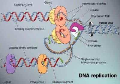
3.8.1 Bacterial DNA Replication 3.8.1.1 Cell Cycle and Replication
DNA, the carrier of genetic information, has to be replicated before Chromosomal DNA replication is linked to the cell cycle by a complex
cellular division takes place (except meiotic division in higher eukarya, regulation system. Initiation, being the critical step, is tightly controlled.
3.9.1). The semi-conservative mechanism uses the parental DNA During the replication reaction, the oriC region (3.8.1.2) of the newly
duplex strands as templates for replication; each parental strand forms formed strand remains unmethylated and the duplex DNA binds to the
a new duplex with a newly synthesized strand. cell envelope. Thus, premature DnaA binding is prevented (which would
otherwise start another replication round). Only later in replication, the
The mechanism of bacterial DNA replication described here applies newly synthesized strand becomes also methylated. Furthermore, the
to many bacterial chromosomes, episomes and plasmids. Unlike concentration of DnaA protein plays an important role for the initiation
eukaryotic chromosomes (3.9.1.2), each of these circular, covalently of DNA replication by initiation of the DNA separation.
3.8.1.2 Initiation of Replication (Fig. 3.8.1-6, Table 3.8.1-2)
The chromosomal replication in bacteria takes place from a single rep-
lication origin (oriC, length 260 bp, Fig. 3.8.1-3) and is mediated by
a single initiator protein, DnaA. This protein interacts specifically with
3.8.1 3 Metabolism 150
five nonpalindromic sequences (DnaA boxes, length 9 bp) at oriC. Four DnaB to form the primosome (at some origins, this is assisted by Pri
of these sites are almost identical and contain the consensus sequence proteins) and synthesizes two leading strand primers.
TTAT(C/A)CA(C/A)A. The left side of oriC is anAT-rich region, consist-
ing of three similar sequences (length 13 bp, each starting with GATC), The DNA directed DNA Polymerase III (Pol III) holoenzyme also
which represent the melting site. FIS protein has a negative effect on the loaded by DnaB is assembled from 10 subunits in a defined order
reaction, HU and IHF proteins, a high ATP concentration (>2 mM), high (Fig. 3.8.1-4). Assisted by the g subunit complex, the dimeric b proces-
temperature (38°C) and transcriptional activation enhance unwinding. sivity factor (b subunit) forms a sliding clamp around the DNA. Pol III*
is finally loaded onto this ring, which clamps to the template strand.
The oriC region also contains no less than eleven 5¢-GATC-3¢ The addition of DNA gyrase completes the formation of the replisome.
boxes, which are target sequences for Dam methylase. Their methyla-
tion is involved in replication initiation. 3.8.1.3 Elongation and Termination (Fig. 3.8.1-6, Table 3.8.1-2)
Gyrase (ATP-dependent topoisomerase type II, breaking and religat-
For initiation, 20 … 30 molecules of DnaA bind to the DnaA boxes ing both strands) travels along the strands ahead of the helicase and
with simultaneous ATP hydrolysis (pre-initiation complex). The DNA introduces negative supercoils into the DNA (underwound state of the
is bent to a loop around the DnaA core. This is supported by binding
of IHF protein (one binding site), and/or FIS protein (four binding
sites) and by unspecific binding of HU protein. It is assisted by simul-
taneous transcription of genes in the oriC region. As a result, strand
separation (melting) of the double helix occurs at the adjacent melting
site, forming an open complex.
Hexameric helicase (DnaB protein) is complexed by accessory
DnaC protein (helicase loader) with simultaneous ATP hydrolysis and
is loaded onto the single-stranded region of the DNA (prepriming com-
plex). The rest of this region is covered by tetrameric single-stranded
binding protein, SSB. Then, the primase enzyme (DnaG) is loaded by
L N RAT-cluster 13mer 13mer R1 IHF M R2 FIS R3 R4
13mer
50bp
AT rich regions DnaA boxes IHF / FIS binding GATC sites Figure 3.8.1-2. Reaction Mechanism of
the DNA Replication
Figure 3.8.1-3. Structure of oriC in Escherichia coli
Table 3.8.1-2. Proteins Involved in Bacterial DNA Replication (Escherichia coli)
Type Protein No. subunits Mol. mass (kDa) Function
Initiator protein DnaA
Integration host factor IHF (heterodimeric) 1 52 binds cooperatively to oriC; promotes double helix opening and DnaB loading
FIS (homodimeric)
Histone-like protein HU 2 (a, b) 11, 11 binds sequence-specifically to double stranded DNA, effects DNA bending
Helicase DnaB
Accessory protein DnaC 2 11 (* 2) binds sequence-specifically to double stranded DNA, effects DNA bending
Primase DnaG
Single strand binding SSB 1 19 binds sequence-unspecifically to double stranded DNA, effects DNA bending
protein
RNA-Polymerase 6 50 (* 6) unwinds parental strands; activates primase on single stranded DNA
Type II topoisomerase
6 29 (* 6) complexes DnaB; delivers DnaB to DNA
1 60 DNA-dependent RNA-polymerase; synthesizes RNA primers
4 19 (* 4) binds sequence-unspecifically, cooperatively to single stranded DNA, stimulates
DNA-Pol III; facilitates DnaB loading
a bb + s 4 (5) 40(* 2), 155,160 + 70 (s)
2 transcriptional activation of initiation; double helix destabilization
2 97 (*2),
Gyrase 90 (* 2) ATP dependent topoisomerase type II, introduces negative supercoils into parental
double helix ahead of the replication fork, removes positive supercoils. Inhibited
DNA Polymerase III subunit a ¸ 1 130 by nalidixic acid and novobiocin.
holoenzyme (core subunit e ˝ core 1 27.5
and t act as dimers subunit q ˛ 1 8.6 catalyzes the main reaction of DNA replication: long strand processivity.
in replication) 1 subunit t 1 71 3¢Æ5¢-exonuclease activity provides proofreading of newly synthesized DNA.
g-complex: g (dimeric), 4 47.5(* 2), 39, unknown, binds e
d, d¢, c, y 37, 17, 15 core dimerization. Gene dnaX encodes also subunit g by translational frameshift
subunit b 2 40.6 (* 2) loads b-subunit to a primed template (‘matchmaker’, ATP-dependent), recognizes
RNA primers at the lagging strand.
DNA Poly- 1 103 processivity factor, ‘sliding ring’, clamps Pol III to primed template, dissociates
merase I 2 easily from rest of Pol III holoenzyme (ÆPol III*).
Ribonuclease RNase H 2 74 Trifunctional enzyme: removal of RNA primers from Okazaki fragments by its
2 36 5¢Æ3¢ exonuclease function, consecutive gap filling by polymerase action and
DNA Ligase Tus 81, 67 proofreading by its 3¢Æ5¢ exonuclease activity. The enzyme also plays a major
topoisomerase IV 177 (* 2) role in DNA repair (3.8.2). Protease splitting yields the ‘Klenow fragment’
Terminator protein MukB (homodimeric) (67 kDa, amino acid residues 324 … 928), which contains the polymerase and the
3¢Æ5¢ exonuclease function. It is widely used in research.
Type II topo- isomerase
removes RNA primer from Okazaki fragments
SMC Protein (structural
maintenance of NAD dependent joining of Okazaki fragments
chromosomes)
inhibits helicase, replication fork arrest
decatenation/catenation, relaxation of supercoiled DNA
chromosome partitioning
1 Terminology of DNA polymerase III: (subunits a + e + q) = core; [(core)2 + (subunit t)2] = Pol III¢; (Pol III¢ + g-complex) = Pol III*; (Pol III* + subunit b) = Pol III
holoenzyme (HE).
2 In E. coli, also DNA polymerase II has been found. This enzyme is likely to be involved in SOS DNA repair (3.8.2.6).
151 3 Metabolism 3.8.1...2
The parental strands are then unlinked by topoisomerases (decate-
nation, 3.9.1.4) and the daughter chromosomes are separated by bind-
ing of MukB and Par proteins (partition).
Figure 3.8.1-4. Assembly of the DNA Polymerase Holoenzyme 3.8.1.4 Fidelity of Replication
The overall error rate is only about 1 per 109 … 1010 nucleotides. This
double helix). This assists the helicase (DnaB) in melting the parental high replication accuracy is due to
helix by separating the strands. The energy is derived from simultane-
ous ATP hydrolysis. Reannealing of the strands is prevented by the SSB • Twofold dNTP base selection mechanism by DNA Pol III at the
protein cover. Then DNA primase (DnaG), activated by helicase, syn-
thesizes RNA primers (length 10 … 12 nucleotides) as starting sequenc- binding and the catalytic step (error rate about 1 per 104 nucleotides)
es for the action of Pol III. The g-complex of Pol III* (see footnote to
Table 3.8.1-2) recognizes the RNA primers and loads a sliding ring of • 3¢Æ5¢-exonuclease activities of Pol III and Pol I removing misin-
dimeric b subunits onto the primer-template junction (ATP dependent).
Thereafter Pol III* moves to this b ring and starts DNA replication (Pol corporated nucleotides (200 … 1000 fold fidelity improvement)
III can, however, carry out the primase reaction itself). Wrongly inserted
nucleotides are removed by the 3¢Æ5¢ exonuclease function of Pol III. • postreplicative mismatch repair (see 3.8.2).
Pol III (like all DNA polymerases) polymerizes dNTPs only in 3.8.2 Bacterial DNA Repair
5¢Æ3¢-direction, therefore only one template strand can be copied
continuously this way (leading strand). The other template strand has 3.8.2.1 DNA Damage
a reverse orientation and can be replicated only discontinuously in The huge DNA molecule can suffer damage in many ways by exo-
5¢Æ3¢-direction by ligating 1.5 … 2 kb long Okazaki fragments (lag- geneous or endogeneous agents. In many cases the bases of DNA are
ging strand, Fig. 3.8.1-5). Thus multiple priming reactions are required. affected. This may cause mutations or interfere with replication and
transcription. The most important damages are:
• Alkylation: Alkylating compounds can modify nucleotides non-
enzymatically. They can be of endogeneous (e.g., S-adenosyl-
methionine) or exogeneous origin (e.g., N-methyl-N-nitrosourea).
Products are, e.g., O6-methylguanine (pairs with thymine!),
3-methyladenine or 2-methylcytosine.
O6-Methylguanine 3-Methyladenine 2-Methylcytosine
Figure 3.8.1-5. Synthesis of Okazaki Fragments The physiological enzymatic methylation of DNA strands, how-
(Lagging Strand, schematically) ever, yields N6-methyladenine, e.g., within the sequence 5¢ GATC
3¢ (in bacteria, 3.8.1) or 5-methylcytosine (in eukarya, 3.9.1),
A b-ring becomes attached to each new primer as described above. which are not subject to repair mechanisms.
Upon completion of the previous Okazaki fragment, Pol III* moves
to this b ring and synthesizes the next fragment until it reaches the • Pyrimidine dimerization: UV Cyclobutylthymidine dimer
primer of the previous one. Then it dissociates from DNA, leaving
the b-ring behind and starts a new synthesis cycle. (The b-ring is later irradiation (l = 200 … 300 nm)
removed by Pol III, too). promotes covalent linking of
pyrimidines (mostly thymine) via
Due to this mechanism, E. coli is able to perform synthesis of both a cyclobutane ring. This distorts
strands with only 10 … 20 molecules Pol III* per cell. The coordina- the DNA structure.
tion of the discontinuous lagging strand synthesis with the continuous
leading strand synthesis is effected by the dimeric structure of Pol • Spontaneous reactions: The most
III (one enzyme for each strand) and by primase activation due to its
association with helicase. frequent spontaneous damage to
DNA is hydrolytic loss of purine
The RNA primer is then degraded by DNA directed DNA poly- bases, leaving apurinic sites.
merase I (Pol I) with 5¢-3¢-exonuclease-function and/or ribonuclease Another hydrolytic reaction is the deamination of cytosine to ura-
H (RNase H). In both cases, the degradation proceeds in the 5¢Æ3¢ cil (U would be read as T in the next semiconservative replica-
direction. Pol I then fills the gap with deoxynucleotides. Finally, the tion!). Deaminations of adenine to hypoxanthine and guanine to
DNA chains are joined by DNA ligase. xanthine occur more rarely. Also demethylation takes place (thy-
mine to uracil).
Termination: Replication terminates in a (non-essential) 0.5 kb termi-
nation region opposite to oriC at the chromosome. At 10 termination • Oxidative damage: Radicals, especially of oxygen (caused e.g., by
(Ter) sites, 10 Tus proteins are bound. Since the Ter sites are non-
palindromic (they do not read identically in both directions), asym- ionizing radiation) can induce unusual modifications of bases:
metric complexes are formed, which block only one of the approach-
ing replication forks by inhibiting strand separation by helicase 5-Hydroxycytosine Thymine glycol 2,6-Diamino-4-hydroxy-5-formamido-
(DnaB). This enables termination of replication at a definite site in pyrimidine
spite of possible speed differences by both forks.
3.8.2 3 Metabolism 152
DEOXYRIBONUCLEIC ACID DEOXYRIBONUCLEIC ACID
PREPRIMING COMPLEX
H PRIMOSOME
GJ
REPLISOME
Figure 3.8.1-6. Bacterial DNA Replication
The newly synthesized DNA is drawn in green
153 3 Metabolism 3.8.2
• Bulky adducts: Compounds which form adducts with bases (e.g., cis- • repair by transfer of sequences from a homologous strand (recom-
platin•1,2-GpG, benzo[a]pyrene•guanine or psoralene•thymine) bination)
cause major distortions of the DNA helix.
The repair systems make use of the redundant information in the DNA
• Double strand breaks: Free radicals induced by ionizing radiation duplex.
or other agents can break both strands of a DNA molecule. This is 3.8.2.2 Direct Reversal of Damage (Fig. 3.8.2-1)
highly hazardous to the cell, since without repair no replication of Reversal of alkylation: Methylation of nucleotides is repaired by
the DNA molecule can take place. DNA repair methyltransferases (e.g., O6-methylguanine-DNA-
protein-cysteine S-methyltransferase). These proteins transfer the
• Replication errors: In spite of proofreading by DNA polymerase methyl group from DNA to a cysteine residue in their active site.
Since this methyl group cannot be removed from the protein, the
(3.8.1.4), occasional errors take place during DNA replication and reaction leads to inactivation of the enzyme (‘suicidal mechanism’).
have to be eliminated by repair systems. Otherwise the fidelity The inactivated enzyme, however, enhances transcription of new
would drop 100…1000 fold. enzyme.
• In heavily damaged cells, the SOS-response (3.8.2.6) tolerates a Photoreactivation: Cyclobutyl pyrimidine dimers are repaired in a
high level of replication errors in order to enable replication at all. photoreactivation reaction by deoxyribodipyrimidine photo-lyase,
DNA repair systems: DNA repair fulfills the vital task of maintaining which catalyzes the cleavage of the cyclobutane ring in a light depend-
the integrity of DNA structure and sequence. In bacteria, several sys-
tems for DNA repair exist, which can be redundant with respect to the ent reaction.
lesion as well as to the implicated proteins. Generally, repair proceeds
by one of the following strategies: The enzyme contains two chromophores [FAD and in different spe-
• direct reversal of damage cies either 5,10-methenyltetrahydrofolate, l ca. 380 nm (E. coli, yeast)
• excision of damage and resynthesis according to the information of max
or 8-hydroxy-5-deazariboflavin, l ca. 440 nm] which absorb light and
the complementary strand (including repair of base pair mismatches) max
initiate dimer splitting by electron transfer to the pyrimidine dimer.
Figure 3.8.2-1. Direct Reversal and Excision DNA Repair Systems in Bacteria (red = damage, green = repair)
3.8.2 3 Metabolism 154
Figure 3.8.2-2. Long patch mismatch repair (Methyl Directed Pathway, E. coli)
(red = newly synthesized strand with mismatch, green = repaired segment)
3.8.2.3 Excision Repair Systems onto a damaged site. After that UvrC binds to UvrB, resulting in the
Base excision repair (Fig. 3.8.2-1): Damage to single bases by oxi- UvrBC-DNA incision complex. In this complex, two incisions are made
dation, deamination, methylation or demethylation (dTÆdU) or base- by UvrC: the first at the fourth or fifth phosphodiester bond on the 3¢
base mispairs are recognized by specific DNA glycosidases, which side of the damage and the second at the eighth phosphodiester bond on
cleave the covalent sugar-base bond, leaving apurinic or apyrimidinic the 5¢ side. UvrD (helicase II) removes the damaged strand. The result-
sites (AP sites). Examples are DNA- (uracil-, 3-methyladenine-, hypox- ing gap is filled by Pol I and the nick is closed by DNA ligase.
anthine- and formamidopyrimidine = 8-hydroxyguanine-) glycosidases.
AP sites may also have risen from spontaneous base losses. AP endo- The transcription-repair coupling factor (TRCF) connects NER
nuclease cleaves the DNA backbone 5¢ of the AP site. Excision exonu- with transcription. TRCF releases stalled RNA polymerase and the
cleases (e.g., DNA deoxyribophosphodiesterase in E. coli) remove the truncated RNA transcript from the damage site and recruits UvrA for
damaged site. The gap is filled in and sealed by DNA polymerase (e.g., consecutive repair as outlined before.
Pol I in E. coli) and DNA ligase. The removal of the damaged site (and
some additional nucleotides) may also be performed by the 5¢Æ3¢ exo- 3.8.2.4 Mismatch repair
nuclease activity of DNA polymerase I. Very short patch repair (E. coli): This system corrects G•T mis-
matches caused by deamination of 5-methylcytosine to thymine in
Alternatively to ‘pure’ DNA glycosidases, those with an associated C(meC)(A/T)GG sequences of fully methylated DNA. The incision
DNA lyase activity carry out additional reactions. Since at the AP site 5¢ of the mispaired T is catalyzed by Vsr protein, an endonuclease.
the hemiacetal deoxyribose ring is at equilibrium with an open-chain Thereafter, DNA polymerase I removes the mispaired T and less than
aldehyde form, the latter can enter a b-elimination reaction leading to 10 more nucleotides by its 5¢Æ3¢ exonuclease activity and fills the
cleavage of the DNA chain 3¢ of the AP site. The biological impor- gap. The nick is then ligated. Mut S and Mut L proteins (see below)
tance of this pathway is uncertain. are also needed, possibly for damage recognition.
Nucleotide excision repair (NER, Fig. 3.8.2-1): Bulky adducts to Long patch repair (Fig. 3.8.2-2): Small insertion or deletion mis-
DNA, as well as damages which cause minor distortions (e.g., pyri- pairs as well as base-base-mismatches are repaired by the methyl-
midine dimers) can be removed by the ABC system, which responds directed mismatch repair (MMR) of E. coli (a related system is the
to these distortions. Hex-dependent pathway of Streptococcus pneumoniae). This system
takes advantage of the fact that for a short time after replication the
The E. coli enzyme activity ‘ABC excinuclease’ is carried out by newly synthesized strand is not yet methylated by Dam-methylase,
the three proteins UvrA, UvrB and UvrC. Dimeric UvrA loads UvrB
Table 3.8.2-1. DNA Repair in Bacteria
System Damage Specificity Implicated Repair Proteins (E. coli) Remarks
“suicidal” mechanism (only one
Reversal of alkylation O6-methylguanine, O4-methylthymidine O6-methylguanine DNA repair methyltransferase I (Ada, methyltransfer catalyzed by one
repairs also O6-methylthymidine and methylphosphotriesters), protein molecule)
O6-methylguanine DNA repair methyltransferase II (Ogt) light dependent (l = 300 … 500 nm)
Photoreactivation pyrimidine dimers deoxyribodipyridine photo-lyase, carrying as chromophores length of repair tract £ 10
methenyltetrahydrofolate + FAD nucleotides
length of repair tract 12–13
Base excision repair uracil, thymine glycols, hypoxanthine, DNA glycosidases, apurinic/apyridinic endonuclease, excision nucleotides
8-oxoguanine, 3-methyladenine exonuclease, DNA polymerase I, DNA ligase length of repair tract £ 10
nucleotides
Nucleotide excision repair bulky adducts, e.g., cisplatin•GpG, UvrA, UvrB, UvrC, UvrD (Helicase II) proteins, DNA length of repair tract ª1000
(Uvr) pyrimidine dimers polymerase I, DNA ligase nucleotides
Very short patch mismatch G•T mismatch (corrected to G•C) in Vsr, MutS, MutL proteins, DNA polymerase I error-prone
repair 5¢-CT(A/T)GG error-prone repair as part of the
SOS regulon, lesion persists
Long patch mismatch repair base-base-mismatch, small insertion/ MutS, MutL, MutH proteins, DNA helicase II, single strand
binding protein, exonuclease I (or exonuclease VII/exonuclease
(methyl-directed) deletion mispairs RecJ), DNA polymerase III holoenzyme, DNA ligase
Recombination repair double strand breaks, distorting DNA sites RecBCD, RecA, RuvC proteins
SOS response DNA polymerase stalled at lesion, DNA RecA, LexA, UmuC, UmuD proteins, DNA polymerase III
single strands remain
155 3 Metabolism 3.8.2
while the parental strand already carries methyl groups at adenine-N6 Its subsequent resolution by RuvC protein brings about exchanges of
in GATC (= Dam) sequences. This differentiates between both DNA the two homologous strands. Several variations of this reaction exist.
strands and insures that sequence information for repair is retrieved
from the unmutated, parental strand. Similar mechanisms (Fig. 3.8.2-4) are used to fill the gap in the daugh-
ter strand opposite a defective site during replication. The defect can
According to the current model, the repair is initiated by the binding be repaired afterwards by the mechanisms described above. However,
of MutS (95 kDa, oligomeric) to the lesion. After the binding of MutL since mismatches are tolerated in the exchange reaction, recombina-
to MutS and MutH to a nearby d(GATC) sequence (up to 1000 bp tional repair is more error-prone than the other repair systems.
away in either direction), an incision is made by MutH (25 kDa) on
an unmethylated DNA strand. Assisted by MutL (95 kDa), the pro- 3.8.2.6 SOS Response (Damage Tolerance Mechanism,
teins form a complex, which incises the non-methylated DNA strand Fig. 3.8.2-5)
5¢ from the GATC site. Subsequently, exonucleases degrade the faulty
strand from here to a point beyond the site of the lesion. If the inci- Agents which cause intense damage to DNA induce a complex emer-
sion is located at the 3¢ side of the mismatch, repair requires the 3¢ to gency repair system in E. coli and similarly in many other bacteria.
5¢ exonucleolytic activity of exonuclease I (ExoI); if the incision is The cells stop dividing and start a special DNA repair mechanism,
located on the 5¢ side, repair requires the 5¢ to 3¢ exonucleolytic activ- sacrificing a high level of fidelity.
ity of either exonuclease VII (ExoVII) or RecJ. The final steps are
performed by SSB, DNA polymerase III and DNA ligase (filling the If the proceeding replication fork meets damaged DNA sequences,
single-strand gap left by the excision). usually in- or post-replication repair mechanisms (as described before)
are initiated. In case of frequent damages, however, DNA polymerase
3.8.2.5 Double-Strand Repair and Recombination III action stops. Thus, single stranded DNA sequences result. RecA pro-
A very difficult situation arises if double strand breaks occur. Similarly, tein (see 3.8.2-5) binds to them and is converted into an activated form,
if during replication a damaged, distorting template sequence is which aids in autoproteolysis of the LexA repressor protein. Under
reached, the replication by DNA polymerase III is interrupted and physiological conditions, this repressor is bound to SOS boxes (con-
reinitiated at some point beyond this site, leaving a gap in the daugh- sensus sequence CTGN10CAG) at more than 20 genes and represses
ter strand. In both cases the strand defects cannot be cured by excision their expression considerably. Removal of this block leads to increased
repair, since no correct template sequence is available in the duplex expression of (among others) UvrA, UvrB and UvrC (see above at NER),
DNA. However, by using the correct sequence from a homologous LexA and RecA proteins as well as of helicase II, DNA polymerase III,
DNA to fill the gap, an intact DNA sequence is obtained, which can be UmuC and UmuD proteins. The latter two enable DNA polymerase
used as a template for damage correction in the other strand (recombi- III to resume replication and to proceed through sites of DNA damage
nation or postreplication repair, Fig. 3.8.2-3). This is possible because despite missing or faulty information from the complementary strand in
fast growing bacterial cells usually start another round of DNA rep- a way not known in detail yet. Although this results in a highly error-
lication before the previous one is completed and cell division has prone DNA replication, it enables some form of replication.
taken place (3.8.1), so that two or more copies of the same gene are
available in one cell. Literature:
In case of double strand breaks, both free 5¢ ends are degraded Friedberg, E.C., Walker, G.C., Siede, W. DNA Repair and
by exonuclease V (RecBCD protein) until a ‘chi-site’ (sequence Mutagenesis. ASM Press, 1995.
5¢GCTGGTGG3¢, spaced at about 10 kb in E. coli) is reached, where
RecBCD switches to a helicase activity. One single-stranded DNA 3¢ Harris, R. S., Ross, K. J., Lombardo, M. J., Rosenberg, S. M. J.
end is helped by the nuclease function of RecA protein (38 kDa) to Bacteriol. 1998;180:989–993.
invade a homologous sequence (Fig. 3.8.2-3). Expansion of the invaded
stretch and gap filling lead to a double Holliday junction (crossover). Messer, W. FEMS Microbiol Rev. 2002;26:355–74.
Modrich, P., Lahue, R. Annu. Rev. of Biochem. 1996;65:101–133.
Sancar, A. Annu. Rev. of Biochem. 1996;65:43–81.
(various authors) Trends in Biochem. Sci. 1995;20:381–439.
Figure 3.8.2-3. Recombination Repair of Figure 3.8.2-4. Recombination Repair of Figure 3.8.2-5. SOS Repair System
Double Strand Breaks Gaps in Daughter Strands
3.8.3 3 Metabolism 156
3.8.3 Degradation of Nucleic Acids The organisms always produce a companion DNA methyltrans-
Nucleases catalyze the cleavage of phosphodiester bonds in DNA ferase, which recognizes the same sequence in endogeneous DNA
(DNases) or in RNA (RNases). DNases play a decisive role during and modifies it immediately after replication by methylation of A or
DNA synthesis, repair and recombination (3.8.1, 3.8.2, 3.9.2), for C residues. This protects the organism’s own DNA from degradation
relief of obstructive superhelical tension as well as in apoptosis (4.3.6, by the restriction enzyme. Thus, the restriction endonuclease and the
7.6) and as a defense measure in restriction of foreign DNA (4.1.4.2). cognate methyltransferase form a restriction-modification (R-M) sys-
RNases are, e.g., of importance in RNA processing (4.1.1.1, 4.2.1.3) tem. Since it cleaves (restricts) infecting DNAs (e.g., viruses) and thus
and also regulate the transcription by degradation of mRNA (4.2). prevents them from parasitizing the cell, the R-M system is also called
Nucleases either remove terminal nucleotides (exonucleases) or act the ‘immune system of the microbes’.
inside of the nucleic acid molecule (endonucleases).
At least four different kinds of R-M systems exist:
3.8.3.1 Exodeoxyribonucleases (Exo-DNases, Table 3.8.3-1)
Exo-DNases are characterized by their cleavage direction, their pref- • Type I enzymes, e.g., EcoK1, carry methylase and nucle-
erence for a single stranded or a double stranded substrate and by
producing mono- or (more rarely) oligonucleotides. In a number of ase activity on the same protein and require Mg++, ATP and
cases, they perform several functions (see below). The nucleases may S-adenosylmethionine for cleavage. They cleave randomly and
dissociate after each catalytic event (non-processive or distributive remote (> 400 bp) from the recognition sequence.
action) or they may remain bound to the polymer until many reaction
cycles are completed (processive or non-distributive action). • Type II enzymes, e.g., EcoR1, recognize mostly palindromic
The reaction scheme of E. coli exonuclease III is schematically nucleic acid sequences and cleave within or near these sequences.
shown in Figure 3.8.3-1. The enzyme is multifunctional: it acts also as (Palindromic sequences repeat each other at the other duplex
an endonuclease specific for apurinic DNA sites (3.8.2.3) and as a 3¢ strand in inversed order, resulting in twofold rotational symme-
phosphatase. Bacterial DNA polymerase I exerts a 3¢ Æ 5¢ exo-DNase try. For examples see Fig. 3.8.3-2.) The enzymes require Mg++ for
activity for proofreading (similarly to eukaryotic DNA Pol d and e) activity. Their homodimeric structure corresponds to the palin-
and additionally a 5¢ Æ 3¢ exo-DNase/RNase function for DNA repair dromic substrate. Many of them generate ‘sticky end’ (‘cohesive
and for removal of Okazaki RNA primers (3.8.2.3; 3.8.1.3). end’) duplex fragments with 5¢ protruding termini (e.g., EcoRI) or
with 3¢ protruding tails (e.g., HhaI). Other enzymes cleave at the
Figure 3.8.3-1. Reaction Scheme of E. coli Exonuclease III center of the recognition sequence and produce ‘blunt end’ frag-
ments (e.g., HaeIII). The cognate methylase is a separate enzyme.
3.8.3.2 Endodeoxyribonucleases (Endo-DNases, Table 3.8.3-2) The cleavage specificity is schematically shown in Figure 3.8.3-2.
The endonucleases often show a strong preference for either single These enzymes are indispensible tools for molecular cloning tech-
stranded or duplex DNA. Endonucleases that function in repair of niques and for DNA sequence analysis. They are named by the
lesions identify the damaged DNA site and incise (nick) the DNA 3-letter abbreviation of the source organism.
at one side of the lesion as a first step towards excision. A second
principal characteristic of these enzymes is the recognition of base • Type IIS enzymes recognize asymmetric sequences of 4 … 7 bp
sequences. A striking example is the cytosine-specific cleavage by T4
endonuclease IV. Pancreatic DNase, E. coli endonuclease I or spleen length. They cleave at a defined distance of up to 20 bp to one side
DNase produce oligonucleotide digests with characteristic sequence of their recognition sequence.
patterns at the 3¢ and 5¢ termini.
• Type III enzymes, e.g., StylT1, have similar characteristics as
Restriction endonucleases occur in a variety of microorganisms. More
than 5000 of them have been identified so far (http://rebase.neb.com/). Type I enzymes, but cleave at specific sites only a short distance
They recognize sequences of 4 … 8 nucleotides in a DNA duplex with (24 … 26 bp) away from the recognition sequence.
extraordinary accuracy and cleave both strands.
Figure 3.8.3-2. DNA Cleavage by Type II Restriction Endonucleases
(® = twofold symmetry axis)
Table 3.8.3-1. Examples for Different Types of Exonucleases
Specificity for 3¢ Æ 5¢ direction Cleavage in 5¢ Æ 3¢ direction either direction
exo-DNase (phage SP3 encoded) E. coli exonuclease VII
Single stranded DNA E. coli exonuclease I, mammalian DNase III exo-DNase (phage l encoded), mammalian DNase IV E. coli exonuclease V (ATP-dependent)
Spleen exonuclease
Double stranded DNA E. coli exonuclease III Yeast RNase
DNA or RNA Venom exonuclease
RNA Exo-RNase II, RNaseQ, RNase BN
Table 3.8.3-2. Examples for Different Types of Endonucleases
Specificity for 3¢ P endproducts Cleavage to 5¢ P endproducts other end products
DNase IV (phage-T4-encoded), yeast DNase Crossover junction endo-RNase (acts only
Single stranded DNA Aspergillus DNase K1 on Holliday junctions)
Type I, II and III restriction enzymes (10.7.2),
Double stranded DNA pancreatic DNase II pancreatic DNase I Bacillus subtilis RNase (yields 2¢,3¢-cyclic
Aspergillus nuclease S1, Mung bean nuclease, phosphates)
DNA or RNA Micrococcal nuclease, spleen endonuclease potato nuclease
Exo-RNase H (acts on RNA : DNA hybrids, degradation
RNA-DNA duplex of Okazaki fragments)
RNase III and RNase P (processing of tRNA and rRNA
RNA RNase T2 = RNase II, pancreatic RNase precursors)
Polynucleotide phosphorylase
157 3 Metabolism 3.8.3, 3.9.1
3.8.3.3 Ribonucleases (RNases, Tables 3.8.3-1 and 3.8.3-2) on DNA replication beyond this point. Here, binding of cyclins
Similarly to DNases, RNases differ by exo- and by endo-activity, (cell cycle regulating proteins, 4.3) to the cyclin dependent kinase
preferences for termini (exo-enzymes) or in some cases for specific Cdc 28 causes activation of transcription factors leading to expres-
sequences (endo-enzymes). Some nucleases even cleave both DNA and sion of genes necessary for DNA replication. Similarly, cell cycles in
RNA. A number of RNase type reactions are not catalyzed by proteins, higher eukarya depend on passage through the restriction or cell cycle
but rather by RNA sequences (ribozymes). E.g., the RNA component commitment point (R) in the G1 phase (4.3.3), where most regulatory
of RNase P catalyzes the processing of untranslated prokaryotic RNA processes leading to cell proliferation take place. After the R point,
(4.1.1.2, as well as some reactions in eukaryota); the protein component cells no longer need growth factors for completion of the cell cycle.
has only assistant function. In a number of cases, eukaryotic group I or
II introns are removed from the rRNA by its own action (self-splicing, In most organisms active genes are replicated early in the S-phase, while
e.g., in Tetrahymena, 4.2). It is speculated that RNA catalysis and self- inactive genes, telomeres, telomere-linked regions and heterochromatin
replication preceded enzyme-protein catalysis during evolution. are replicated later on. The influence of various factors like histones,
nucleosomes, the nuclear matrix and matrix associated enzymes (e.g.,
Literature DNA topoisomerase II, histone acetyltransferases, histone deacetylases,
Lin, S.M., Roberts, R.J. (Ed.) Nucleases. CSH Monograph Series, 1985. poly-ADP-ribosyltransferase etc.) as well as the unfolding mechanism
Kornberg, A., Baker, T.A. DNA Replication. 2nd Ed. Freeman, 1992. of the densely packed chromatin are very complex and will not be dis-
Roberts, R.J., Macelis, D. Nucl. Acid Res. 1996;24:223–235. cussed here.
3.9 Nucleic Acid Metabolism in Eukarya 3.9.1.2 Initiation of Replication (Fig. 3.9.1-2, Tables 3.9.1-1
and 3.9.1-2)
Helmut Burtscher
Initiation of replication takes place at origins or initiation zones. These
3.9.1 Eukaryotic DNA Replication DNA regions are recognized by specific proteins or protein complexes
With the exception of the second meiotic division of germ cells, DNA which catalyze localized unwinding and load replication proteins onto
replication is a necessary prerequisite of cellular division. DNA must the exposed single strands. The order of assembly of replication fac-
be replicated with high fidelity to ensure correct information transfer tors is highly conserved across all species.
to daughter cells. In eukarya several replicative DNA polymerases,
some of them with intrinsic exonuclease activity, are involved in this Unlike bacterial chromosomes, eukaryotic chromosomes contain
extremely accurate process. multiple origins of DNA replication. On a typical mammalian chro-
mosome these are spaced 50 … 100 kb apart. Not all replication ori-
Although in eukarya it is actually chromatin that replicates (the gins are activated simultaneously. Some origins are used early, some
complex of DNA, histones and other proteins, attached to the nuclear late in S-phase and not all seem to be utilized in every cell or during
scaffold, 2.6.4), the following section concentrates on the replication every cell cycle. Early replicating origins seem to be associated with
of DNA, which proceeds in a series of integrated protein-protein and transcriptionally active regions whereas late replicating origins are
protein-DNA interactions and enzymatic reactions. located near repressed regions. However, it looks as if it is not chro-
matin accessibility or pre-replication complex formation but a later
3.9.1.1 Cell Cycle and DNA Replication step that determines origin firing.
DNA replication must be precisely linked to the cell cycle (summa-
rized in Fig. 3.9.1-1). It depends on the passage through a special Re-initiation from the same origin during a single cell cycle must
point in the G1-phase, at which cells commit to another round of repli- be blocked by all means. This is ensured by redundant mechanisms,
cation and cell division (see below). DNA is replicated exclusively in many if not all of which are regulated by cyclin-dependent kinases.
the S-phase. For the next round of replication, a passage through the
M phase must normally have taken place. Table 3.9.1-1. Replication of Genomes
The mechanisms underlying these controls are still being eluci- Species Size (bp, Number of Number of Replication Duration of Genome
haploid)
Figure 3.9.1-1. Phases of the Mammalian Cell Cycle Chromosomes Origins (total) Replication
Yeast 1.4 * 107
dated. More details of the regulatory aspects are given in Section 4.3. 16 250 … 400 30 min
Human 3 * 109
• The chromatin must be in a replication competent state: A pre- 2 * 22 + 2 20,000 … 50,000 8h
replication complex of DNA and ‘licensing factors’ (3.9.1.2) must In yeast (S. cerevisiae), replication origins are called autonomously
have been formed, which dissolves during the later phases of the cell replicating sequences (ARS). A stretch of 100 to 150 nucleotides is
cycle and thus prevents entering an untimely round of replication. required for maximal function, arranged in domains A and B. Domain
A contains an essential ARS consensus sequence, 5¢-(A/T)TTTA(T/C)
• Also, external signals (e.g., nutrient supply in unicellular organisms (A/G)TTT(A/T)-3¢. Origins in yeast resemble promoters (3.9.2).
Replication enhancer sequences can be located at distances up to 1 kb
or hormonal messages in multicellular organisms) are required to on either side of the start position and in both directions.
initiate DNA replication and cellular division. In yeast, coordination
of cell growth and DNA replication occurs at a unique point in the A complex of six essential proteins in yeast (origin recognition com-
G1 phase of the cell cycle, which is called START (Fig. 3.9.1-2). plex, ORC, homologues in other eukarya) binds in ATP-dependent
This control step irreversibly commits the cell to another round of manner to ARS and remains there throughout the cell cycle. The acti-
cell division, as most external cell cycle regulators have no effect vating proteins Cdc6, Cdt1 and a complex of Mcm proteins (‘licensing
factors’) become available during mitosis (when the nuclear mem-
brane dissolves) and are part of the pre-initiation complex at the end of
M-phase, which is essential for initiation of replication. Cdt1 recruits
Cdc45 into the complex. Thereafter, Cdc6 and Cdt1 leave the complex
and are either degraded or sequestered for most of the cell cycle (The
Mcm complex moves along with the replication fork). Complex forma-
tion and dissolving is controlled by the cyclin/cyclin dependent kinase
complexes Clb1, 2, 3, 4-Cdc28 and Clb5, 6-Cdc28, respectively (4.3.2).
Identity and structure of replication origins in higher eukarya are
probably similar but more complex. Initiation sites can be distributed
over broad regions (initiation zones) of several kb length possibly due
to more complex regulation requirements. The chromosomal structure
seems to be a major influence. Today, methods are available for isola-
tion of active origins, e.g., from CHO and other cells.
3.9.1 3 Metabolism 158
3.9.1.3 DNA Polymerases (Pol, Table 3.9.1-3) polymerases which are non-processive and copy DNA with low
Eukarya have an amazing number of DNA polymerases, e.g., at catalytic efficiency in an error-prone but nevertheless essential
least 14 DNA-dependent DNA polymerases are encoded in the manner.
human genome. These polymerases are grouped in families based
on sequence homologies in their catalytic domains; they can also In yeast three DNA polymerases (a, b, e) are essential for DNA
be grouped according to function: Replicative polymerases which replication. Their sequences are highly conserved (human and yeast
copy the genome with high fidelity and trans-lesion or bypass DNA polymerase a show 31 %, bovine and yeast DNA polymerase d
44 % homology).
External Trigger
START
Cdt Cdt
1 1
Mcm (2-7) Mcm (2-7)
Cdt
1
Figure 3.9.1-2. Model of Replication Initiation in Yeast
(Spatial Arrangements of the Factors are unknown)
Table 3.9.1-2. Essentials for Initiation of DNA Replication in Yeast (Saccharomyces cerevisiae)
Type DNA Sequence/Protein Mol. mass kDa Function
Factor
Origin DNA element ARS Autonomously replicating sequence, core (A site ) + 3 adjacent B sites
DNA element Abf binding sequence 3¢ or 5¢ from core ARS, consensus sequence (A/G)TC(A/G)(T/C)NNNNNACG
DNA elements MCB/SCB Cell cycle boxes, located in the promoter of genes transcribed in late G1 phase, bound by
transcription factor Mbf and Sbf, respectively
Proteins:
Origin recognition ORC (subunits: Orc 1…6) 104, 71, 72, 61, Remains bound to ARS (core + 3 sequence) throughout the cell cycle
complex 55, 50
Activating proteins (in nucleus Cdc6 58 Activates ORC/ARS, required for binding of Mcm proteins and recruitment of Cdc45
74 Activates ORC/ARS, required for recruitment of Mcm-complex
only in M + G1 phases) Cdc45 68 Licensing factor, associates with Cdc6 to enable association of mcm proteins with chromatin
Cdt1
Mcm proteins (in nucleus only Mcm2 99 Activates ORC/ARS, possibly ‘licensing factor’
108 Binds 3¢ to ARS, activates ORC/ARS, opens duplex (helicase?).
in G1 and S phases) Mcm3
Analogy in humans: P1 = DNA Pol-primase accessory factor, targets Pol a?
also: Mcm4 (Cdc54), Mcm5 (Cdc46), Mcm6, Mcm7
(Cdc47)
Cyclins G1 type: Cln1, 2, 3 62, 62, 65 G1 phase cyclins, associate at START with kinase Cdc28, activate Mbf/Scf
Protein kinases B type: Clb5, 6 50, 44 Late G1 and S phase cyclins, associate with kinase Cdc28, inhibited by Sic1
Clb1, 2, 3, 4 55, 56, 49, 54 G2 and M-phase cyclins
Cdc28 (cyclin-dependent, 34 a) Activation at START after association with G1 cyclins Cln1, 2, 3, budding
present throughout cell b) Association with B cyclins Clb5, 6: phosphorylates Cdc7, Swi6, Cdc10, RF-A
cycle) Generally: causes DNA synthesis, spindle formation
Activating protein Cdc7 58 Expression depends on Mbf1 bound to MCB sequence, becomes activated by Dbf 4,
Dbf4 phosphorylates Orc or other proteins in late G1 phase
80 Targets Cdc 7 protein kinase to DNA
Inhibitor Sic1 (p40) 32 Inhibits Cdc28–Clb5, 6 complex, degraded after phosphorylation by Cdc28/Cln
Ubiquitin-protein ligase Cdc34 34 Ligates Sic1-P with Ub for degradation, thus activates Cdc 28/Clb5,6-complex
Transcription factors Abf1 81 ARS binding factor, enhancer, required for origin function
Mbf (subunits: Mbp1, Swi6) 94, 91 Bind after phosphorylation to MCB and SCB DNA sequences, respectively, start transcription of
and Sbf ribonucleotide reductase, thymidylate synthase, Pol a-primase, Clb5, 6, Cdc6, 7, Cdc46, Dbf4,
Orc6 etc.
Polymerase / primase and replication factor RF-A See 4.2
159 3 Metabolism 3.9.1
Table 3.9.1-3. Replicative Eukaryotic DNA Polymerases
Polymerase Source Number of Mol. mass kDa Function
subunits
Initiation of both strands. Subunit p68 is periodically phosphorylated in a cell-cycle dependent manner.
a (I) yeast 4 167, 79, 62,48 Tightly associated primase activity. Starts Okazaki fragment synthesis. Synthesizes an RNA primer of
b (IV) (primase) 8 … 10 nt length, extends it with an initiator DNA of less than 50 nt. No exonuclease activity.
human 4 180, 68, 58, 48
yeast 1 Involved in base excision repair (3.9.2.3). Essential in mammals (as shown in mice), this enzyme is mutated
human 1 68 in up to 30 % of human cancers; not essential for viability in yeast.
39
Replication of mitochondrial DNA, similar enzyme in chloroplasts, human enzyme consists of a homodimer
g (m) yeast 1 144 of accessory subunits and a catalytic subunit. The enzyme is encoded in nuclear DNA.
d (III) human 3 140, 55 (2)
Synthesizes leading strand and completes Okazaki fragment synthesis. No primase activity. Requires PCNA
yeast 3 125, 55, 40 (Table 3.9.1-5) for binding to DNA. Long strand processivity. Contains 3¢ Æ 5¢ exonuclease for proofreading
human 4 125, 66, 50, 12 (by removing mononucleotides). Also involved in DNA-repair and recombination. Essential.
e (II) yeast 5 255, 80, 34, 30, 29 Similar to Pol d. Long strand processivity even without presence of PCNA. 3¢ Æ 5¢ exonuclease activity,
human 4 258, 59, 17, 12 degrades to 6 … 7 nt long oligonucleotides. Participates in DNA replication (leading strand) and repair.
Essential.
3.9.1.4 Replication Forks (Fig. 3.9.1-6) strand) by ligating short Okazaki fragments (about 100 … 200 nucle-
The general aspects of DNA replication are discussed in Chapter 3.8.1. otides in eukarya), which are also synthesized in 5¢Æ3¢ direction.
Eukaryotic replication forks move with a speed of about 30 … 50 nt/ Leading and lagging strand polymerases presumably act in concert,
sec. (in vitro, on ‘naked’ DNA, about 500 nt/sec. are observed). The resulting in the periodic formation of unreplicated looped lagging
large number of replication origins shortens the replication time of strand DNA (trombone model). Although more effort seems to be
mammalian chromosomes from more than 1000 hours (calculated for needed for lagging strand processing, both strands are synthesized at
a single origin) to ca. 8 hours. identical rates.
Although it is still generally assumed that the replication machin- The Okazaki fragments are started by RNA primers and initiator
ery travels along the DNA, there are also indications for a relatively DNA. The Pol a /primase performing these reactions is consecutively
immobile replication apparatus, where the DNA fork is being threaded replaced via a polymerase switch by Pol d, which continues replica-
through. (Analogous possibilities also exist for transcription complexes, tion until the previous Okazaki fragment is reached. Removal of the
4.2.1.2). RNA primers requires the combined action of the 5¢Æ3¢ exonuclease
MF1 and RNase H. The resulting gap is filled by a DNA polymerase
After the initiation phase (which is presented in Section 4.3.2 for and closed by DNA ligase I.
yeast), helicases unwind the DNA double strand and DNA topoi-
somerase I relieves the positive superhelical tension (Fig. 3.9.1-3, There is still some uncertainty about the detailed mechanism of
one strand is nicked and released after passing past the other strand). DNA replication. In particular, the exact spatial arrangement of the
Both reactions are energized by ATP hydrolysis. DNA directed DNA DNA replication system is still unclear. Several models have been
polymerase a/primase (Pol a /primase) attaches itself to the single proposed which differ in various aspects. Therefore, Figure 3.9.1-6
strands, the still open portion of which is covered by RPA (= RFA). will probably not be exact in all details.
The primase synthesizes a short stretch of RNA complementary to
the template DNA, which is extended as initiator DNA (ca. 34 nt) by The completed DNA strands wind around histones and are further
Pol a. Then a polymerase switch takes place: RF-C displaces Pol a compacted, using scaffolding proteins (2.6.4). The relaxing of negative
by binding to the 3¢ end of the initiator DNA. It then recruits PCNA supercoil tensions, which occur during these reactions, is achieved by
to form a trimeric ring around the DNA strand, which enables DNA DNA topoisomerase II. Another function of this enzyme is the removal
directed DNA polymerase d (Pol d) to join and to continue DNA repli- of ‘knots’ of the DNA strands (decatenation). The enzyme breaks both
cation. PolD is able to proofread the newly synthesized DNA strands. DNA strands and guides them through another DNA duplex.
Analogous to bacteria (3.8.1.3), replication of one template strand Eukaryotic topoisomerase type IB (Fig. 3.9.1-3) acts preferentially
can proceed continuously in 5¢Æ3¢ direction (leading strand as on double stranded DNA. A tyrosine hydroxyl group of the enzyme
described above). Since the other template strand has a reverse orien- temporarily forms a covalent bond with the 3¢ phosphate group of one
tation, its replication can be performed only discontinuously (lagging of the DNA strands. After the free 5¢ end has turned in order to relax the
twisting stress, release of the nick takes place. No supply of additional
Table 3.9.1-4. Eukaryotic DNA Polymerases With Special Functions
Polymerase Source Number of subunits Mol. mass Function
kDa
h yeast 1 72 Bypass of UV-induced lesions, accumulates at replication forks after DNA damage; defects cause xeroderma
78 pigmentosum variant disease
human 1
Bypass of normal replication-blocking DNA lesions
i yeast 80
human 1 Bypass of normal replication-blocking bulky DNA lesions
99
k yeast
? Base-excision repair, non-homologous end joining, has DNA polymerase and terminal transferase activities
human 1 63
l yeast ?
human 1
μ yeast ? ? Non-homologous end joining, sister-chromatid exchange
55 DNA repair, e.g., interstrand DNA crosslinks (?), ATPase activity associated
human 1 Bypass of normal replication-blocking DNA lesions
?
q yeast ? 290
human 1 173, 112, 29
353, 24
z yeast 3
human 2
Revy yeast - - (in yeast part of POLZ)
n human 1 138 Incorporation of dC opposite abasic sites
yeast ? ? unclear
human 1 100
3.9.1 3 Metabolism 160
Table 3.9.1-5. Other Important Proteins Involved in DNA Replication
Enzyme Source No. subunits Mol. mass kDa Function
DNA Helicases eukarya 1 46…110 Associate with various DNA (and RNA) polymerases and catalyze DNA unwinding by disruption
of hydrogen bonds between duplex DNA strands (ATP dependent). Action proceeds either in
5¢Æ 3¢ or in 3¢ Æ 5¢ direction, yielding stretches of single stranded DNA for replication, repair
(3.9.2), recombination, transcription and conjugation. More than 20 eukaryotic helicases have been
characterized, their role and mechanism of action still remains to be elucidated in most cases.
DNA eukarya 1 95…165 Removes positive superhelical tension by inducing a transient single strand nick into the DNA template
Topoisomerase I ahead of the replication fork. Inhibited by camptothecin.
RPA (RFA) yeast 3 69, 36, 13 Unspecific single strand and sequence-specific double-strand DNA binding protein. The 36 kDa subunit
mammals 3 70, 34, 11 is periodically phosphorylated by a cyclin-dependent kinase during the cell cycle. The 69 kDa subunit
RFC contains single strand DNA binding and origin-unwinding activity.
yeast 4 120, 40, 37, 36
Proliferating cell mammals 3 140, 41, 37 Essential accessory factor to DNA polymerases d and e. It binds to the primer terminus (ATP dependent)
nuclear antigen and recruits PCNA as a ring around DNA. The subunits are highly homologous among each other and
(PCNA) eukarya 3 (homotrimeric) 26 (*3) between eukarya. Possibly analogous to the complex of E. coli DNA Pol III (3.8.1.2).
Ribonuclease H yeast 1 118 Essential accessory factor to DNA Pol d. Binds possibly to DNA analogous to the b-subunit of E.
Exonuclease MF1 human 1 43 coli DNA Pol III (3.8.1.2). DNA is thought to be threaded through a ring (sliding clamp) of PCNA,
yeast 1 (Cdc9) ca. 85 preventing DNA polymerase to dissociate from the DNA during synthesis. Participates in nucleotide
DNA ligase mammals 1 (Ligase I) 102 excision repair of UV-damaged DNA (3.9.2.2).
yeast many 103 (cat. subunit)
Telomerase human many (>9) 127 (cat. subunit) Degradation of RNA primers in Okazaki fragments.
eukarya 2 (homodimeric) 100…180 (*2)
DNA Degradation of RNA primers in Okazaki fragments in 5¢Æ 3¢ direction. Cooperates with RNase H.
Topoisomerase II eukarya 2* (2a, 2b, 3, 4) 14, 14, 15, 11
Histones Catalyzes the formation of phosphodiester bonds at single-strand breaks in double-stranded DNA. Of
the 3 mammalian DNA ligases, ligase I is used in replication.
RNA dependent DNA polymerase, contains essential RNA. Extends 3 DNA strand ends by addition of
telomeres.
Required for segregation of daughter DNA molecules after replication. Relaxes supercoils; acts via
cutting and re-ligating of double strands. It is also important as a nuclear scaffolding protein.
Basic chromosomal proteins, essential components of nucleosomes. Histone H1 binds to DNA between
nucleosomes (2.6.4)
energy is required. (Contrary to this, in bacterial topoisomerase type IA protein configuration, which allow the passage. [A similar mechanism
the 5¢ phosphate group is temporarily bound by tyrosine.) is used by bacterial gyrase (3.8.1.3), where a DNA duplex of about
140 bp is wrapped around the enzyme, crossing itself. Duplex strand
The dimeric topoisomerase type II (Fig. 3.9.1-4) breaks both passage through the gap produces negative supercoils.]
strands of duplex DNA 4 bp apart. A covalent bond between 5¢ phos-
phate groups of the DNA and a pair of tyrosyl groups of the enzyme 3.9.1.5. Telomeres (Fig. 3.9.1-5)
is formed. Then another unbroken DNA duplex passes through the In replicating the lagging strand of linear DNA a problem arises, when
gap. The reaction requires ATP in order to achieve the changes in the the 3¢ end of the template strand is reached. The last RNA primer can-
not be replaced by DNA since there is no further template for synthe-
Figure 3.9.1-3. Mechanism of DNA sizing another primer/Okazaki fragment. Therefore, with every round
Topoisomerases IB (3’, Rotation Model) of replication, shortening of DNA would occur. In order to avoid grad-
ual loss of essential genes, linear DNA carries telomere extensions
of up to 2000 short, tandemly repeated DNA sequences at its 3¢ end
(species specific, 5 to 8 bases long, in humans TTAGGG).
In vitro, the G-rich sequences frequently fold back onto them-
selves by non-Watson-Crick G-G base pairing, forming hairpin loops
Figure 3.9.1-4. Mechanism of DNA Topoisomerases II Figure 3.9.1-5. Formation of Telomeres
161 3 Metabolism 3.9.1
quiescent in G0 phase
DEOXYRIBONUCLEIC ACID S phase
M phase/G1 phase 'PREPRIMING
COMPLEX'
'PRIMOSOME'
(3.9.1.5)
'REPLISOME'
Figure 3.9.1-6. Eukaryotic DNA Replication (Not all details covered)
(t-loop) and covering the strand ends. This way, telomeres might keep DNA from RNA templates, it resembles reverse transcriptase (5.4.1,
chromosomes from fusing end-to-end, thus preventing chromosome except for using an internal template). The telomeres are bound by
breakage and loss during cell division. Chromosomes lacking telom- various telomere binding proteins (TBPs, like POT1, TRF1, TRF2)
eres are not stably maintained. which play an important role in telomere length control.
The formation of these extensions is catalyzed late in S phase by While lower eukarya like yeast and also the germ line cells of
telomerase. The enzyme contains an essential RNA component serving higher eukarya contain telomerase, most somatic cells of higher
as template for the synthesis of the telomere repeats. By synthesizing eukarya do not express this enzyme (fetal cells, blood stem cells and
3.9.1...2 3 Metabolism 162
many mouse cells seem to be an exception). Therefore telomeres can arrest cell cycle progress at defined “checkpoints” when damaged
shorten with every round of DNA replication in somatic cells. This DNA is detected (4.3.6).
might limit their life-span and be part of a molecular clock measur-
ing the age of cells. Aberrant telomerase activity has been implicated Figure 3.9.2-1 gives an overview of the basic features of different repair
in the loss of normal cellular function and seems to be an inherent pathways. Covalent modifications (ubiquitinylation, SUMOylation, etc.)
property of malignant cells making telomerase a promising target which also play an important role in many of these processes have been
for tumor therapy. omitted.
3.9.1.6 Fidelity of Replication 3.9.2.2 Direct Reversal of Damage (Fig. 3.9.2-1)
Accurate replication is essential for maintaining genome stability. Reversal of alkylation: Homologues to the specialized alkyltrans-
Base selectivity alone would lead to more than 1 error for each 100 ferases of E. coli for direct repair of alkylation damage (3.8.2.2) have
nucleotides polymerized. Additional mechanisms, like proofreading also been found in eukarya. O6-methylguanine-DNA-protein-cysteine
and post-replicative mismatch repair (3.8.2.4) reduce the overall error S-methyltransferase (O6-MGT I) is a ‘suicide’ enzyme that removes
rate to about 1 per 1010 nucleotides. This rate, as well as the relative methyl groups from O6-methylguanine to a cysteine-SH group of the
contributions of the mechanisms for enhancing fidelity are similar to enzyme at the expense of its own inactivation.
bacteria (3.8.1.4). The most frequent error is the deletion of a single
nucleotide. Among the replicative DNA polymerases pol e is the most Photoreactivation: A deoxyribodipyrimidine photolyase (similar to
accurate eukaryotic DNA polymerase, while pol a is the least accurate bacteria, 3.8.2.2) has been found in S. cerevisiae, some other fungi
one (it lacks proofreading ability). In some hereditary forms of human and in algae, but so far not in mammalian cells. A similar enzyme in
cancer, impaired mismatch repair leads to several thousand-fold higher animals repairs the 6-4 photoproduct (another pyrimidine dimer).
error rates in DNA replication.
3.9.2.3 Excision Repair Systems (Fig. 3.9.2-1)
Literature: Excision repair relies on the redundant information in the DNA duplex
to remove a damaged base or nucleotide and to replace it with the cor-
Aparicio, O.M. et al: Cell 1997;91:59–69. rect base by using the complementary strand as a template.
Bell, S.P., Dutta, A. Annu. Rev. Biochem. 2002;71:333–374.
Bloom, J., Cross, F. R. Nat. Rev. Mol. Cell Biol. 2007;8:149–160. Base excision repair (BER): With this mechanism, damages caused
Diller, J.D., Raghuraman, M.K. Trends in Biochem. Sci. by ionizing radiation, oxidizing and methylating agents (including
products of the endogenous metabolism) and by loss of bases are
1994;19:320–325. repaired. It has only a limited substrate range since damaged bases
Loeb, L.A., Monnat, R.J. Nat. Rev. Genetics 2008;9:594–604. must be recognized by specific DNA glycosylases.
Roca, J. Trends in Biochem. Sci. 1995;20:156–160. In base excision repair single altered bases are targeted. Each of the
DNA-glycosylases (e.g., uracil DNA-glycosylase or 3-methyladenine
Sugino, A. Trends in Biochem. Sci. 1995;20:319–323. DNA-glycosylase; the EC list uses ‘glycosidase’) recognizes specifi-
cally a particular type of modified bases and removes them by hydro-
Stillman, B. Cell 1994;78:725–728. lyzing the N-glycosidic bond between the base and the deoxyribose-P
backbone of the DNA. The resulting ‘apurinic’ or ‘apyrimidinic’ site
Toyn, J.H., et al. Trends in Biochem. Sci. 1995;20:70–73. (AP site) is recognized by specialized AP-endonucleases which cut
the DNA backbone 5¢ (upstream) to this location. A deoxyribophos-
Wang, T.A., Li, J.J. Current Opin. in Cell. Biol. 1995;7:414–420. phodiesterase (dRPase, an exonuclease) removes the base-free sugar
phosphate. DNA polymerases (b, possibly also a or e) fill the gap with
Wang, J.C. Ann. Rev. of Biochem. 1996;65:635–692. the correct nucleotide. Then the strand is closed by action of a DNA
ligase. In mammals, this is DNA ligase III.
Wang, J.C. Nature Rev.Mol.Cell Biol. 2002;3:430–440.
Nucleotide excision repair (NER): This mechanism is coupled to
3.9.2 Eukaryotic DNA Repair transcription with selective preference to the transcribed strand. Since
NER responds to distortions in DNA structure and not to individu-
3.9.2.1 DNA Damage and Principles of Repair ally recognized lesions, it is an almost universal repair system which
DNA is a highly vulnerable molecule due to its enormous size. It can removes a wide spectrum of structurally unrelated damages.
be damaged by many exogenous agents (e.g., environmental chem-
icals, UV or ionizing radiation). Also, spontaneous DNA damage Examples are most UV-induced photoproducts (e.g., cyclobutane
arises from hydrolytic removal of bases, deamination of cytosine pyrimidine dimers, 3.8.2.1), bulky chemical adducts (e.g., benzo[a]
to uracil and (more rarely) adenine to hypoxanthine and guanine to pyrene•guanine adducts caused by smoking, cis-platin•guanine
xanthine, non-enzymatic DNA methylation by S-adenosyl methio- adducts), methylated bases (e.g., O6-methylguanine) and intrastrand
nine or damage by free oxygen radicals (reactive oxygen species, as well as interstrand crosslinks. However, sometimes it also acts
ROS) (3.2.5.8, 3.8.2.1) as well as replication errors. In humans, (inefficiently) on mismatches with no strand preference, which can
these types of spontaneous damage occur at a daily rate in the order lead to fixation of mutations.
of 50,000 per cell.
Although there are no sequence homologies to the E. coli UvrABC
The ability to minimize the number of inheritable mutations endonuclease system (3.8.2.3), the DNA strand in eukarya is cut simi-
caused by damage to DNA is essential for all organisms in order to larly on both sides of the lesion by a specialized excision nuclease
maintain genome integrity. Besides mutagenicity, DNA damage can complex. Eukaryotic nucleases hydrolyze the 3rd to the 8th phosphodi-
also interfere with transcription and replication. Therefore, an intri- ester bond 3¢, followed by the 20th to 25th phosphodiester bond 5¢ of
cate network of various systems for recognition and repair of DNA the lesion, removing a patch of 22 to 32 (in humans 27 to 29) nucle-
damage has evolved. otides. The incision step alone requires many different proteins (10 in
yeast, 16 in humans). A selection is shown in Figure 3.9.2-1 and Table
DNA repair in eukarya appears to follow the same basic reac- 3.9.2-1. RFA temporarily covers the separated strands. The gap is then
tions as in bacteria (3.8.2), but is more complicated due to the larger filled by DNA polymerase d or e and the continuity of the phosphodi-
genome and its more complex structure. Similarities in DNA repair ester backbone is restored by DNA ligase.
mechanisms extend from yeast to humans. Some of the components
can even be exchanged in vivo. Some of the repair factors in humans are also part of the transcription
machinery (4.2), e.g., TFIIH, TFIIJ. Coupling of repair to transcrip-
Repair is often associated with DNA replication and transcription tion is effected by CSA/ERCC8 and CSA/ERCC6 proteins, which
when DNA is supposedly more accessible and repair is more essential temporarily replace RNA polymerase stalled by the NER system.
than in a quiescent state. Transcriptionally active regions are repaired
faster than ‘silent’ regions (transcription-coupled repair). Genes tran-
scribed by RNA polymerases I or III are not subject to this specific
type of DNA repair. Eukaryotic cells have checking mechanisms that
163 3 Metabolism 3.9.2
Reversal of alkylation Base excision repair (similarly:
short patch mismatch repair)
DEOXYRIBONUCLEIC ACID
UV
hν
Photoreactivation
Nucleotide
excision
repair
Figure 3.9.2-1. Systems of Eukaryotic DNA Repair. The terminology of the human system is used.
Most of the activities first described in E. coli and in S. cerevisiae 3.9.2.4 Mismatch Repair
have also been found in mammalian cells. Base pairs in a double-stranded DNA helix not conforming to the
Watson-Crick rules (A-T, G-C, 2.6.1) are called mismatches. Such
Table 3.9.2-1. Protein Factors in Eukaryotic Nucleotide Excision Repair incorrect base pairs arise for several reasons:
(Selection)
• occasional errors in proofreading mechanisms during DNA-
Gene product Mol. mass kDa Function
(human) replication
Damage excision: • formation of incorrect heteroduplexes between DNA-strands dur-
XPA 31 recognition of damage (zinc finger) ing recombination
RPA (RFA) (see 3.9.1.4) same, binds to single-stranded DNA • deamination of 5-methylcytosine to thymidine, causing a G-T
XPC/RAD23B 102, 43 stabilization of preincision complex mismatch.
TFIIH (see 3.9.1.4) formation of preincision complex, transcription- Thus, this type of repair system is preferably connected to replica-
repair coupling tion and recombination events. It follows the same principles as the
excision repair systems mentioned above. The main problem is the
XPB (ERCC3) 89 3¢Æ5¢ helicase, TFIIH subunit distinction of the correct from the incorrect base, since both molecules
are normal constituents of DNA. As in E. coli, mismatches in mam-
XPD (ERCC2) 87 5¢Æ3¢ helicase, TFIIH subunit malian cells can be repaired by two different mechanisms depending
on the sequence context:
XPG (ERCC5) 133 DNA incision, 3¢ side of lesion
• A short patch system with preference for G-T mismatches originating
XPF (ERCC4)/ 104, 33 (1:1 DNA incision, 5¢ side of lesion
from deamination of (5-methyl-)C at CpG sites. It resembles base
ERCC1 complex) excision repair (3.9.2.3) and likewise shows restricted specificity.
After thymine removal, 10 or less nucleotides (down to a single
Gap filling:
RFC (see 3.9.1.4) loads PCNA onto DNA (ATP-dependent)
PCNA, DNA polymerase b, d or e, DNA ligase – see 3.9.1.3
3.9.2, 3.10.1 3 Metabolism 164
one in mammals) are excised. The resulting gap is closed by DNA 3.9.2.7 DNA Repair and Human Diseases
polymerase b. Defects in DNA repair cause severe diseases, e.g., most human cancers
result from DNA damage. Human DNA repair has mostly been studied
• Likewise, the mismatch-specific thymine-DNA-glycosylase of in genetic diseases with impaired repair function. Examples are xero-
derma pigmentosum (high sensitivity against UV radiation), Fanconi
humans is also able to remove dU generated at CpG sites by deamina- anemia (cross-link sensitive), Cockayne’s syndrome and trichothiodys-
tion. There are also hints for a mismatch repair mechanism in mam- trophy, all of which are caused by defects in nucleotide excision repair.
mals which removes A (mispaired to G) by incisions at both sides. In ataxia telangiectasia the repair of double-strand breaks is defective;
the patients are extremely sensitive to ionizing radiation. Damage in
• a long patch mechanism using MutS and MutL homologues simi- genes for long patch mismatch repair mechanisms (hMSH2 and hPMS1
on chromosome 2, hMLH1 on chromosome 3p21 or hPMS2 on chro-
lar to the bacterial methyl-directed pathway (3.8.4, recognition of mosome 7) are associated with hereditary nonpolyposis colorectal
the correct strand at methylated moieties after replication, or of the cancer (HNPCC; Lynch syndrome II, Muir-Torre syndrome), which is
defective strand at breaks). This system detects mismatches in many characterized by instabilities of simple repeated sequences. This is one
different contexts. Fairly large pieces of DNA (up to >1000 bases) of the most common genetic diseases of humans (1 in 200 individuals).
are excised, this may occur in either direction. The gap filling is simi-
lar to nucleotide excision repair, possibly by DNA polymerase d or e. Literature:
3.9.2.5 Double-strand Repair and Recombination Bergink, S., Jentsch, S. Nature 2009;458:461–467.
One of the severest DNA lesions occurring in a cell is a double-strand Branzei, D., Foiani, M. Nat. Rev. Mol. Cell Biol. 2008;9:297–308.
break, because it completely disrupts the integrity of the DNA molecule. Friedberg, E.C., et al. DNA Repair and Mutagenesis. 2nd Ed. ASM
This is the most frequent effect of ionizing radiation. Double strand ends
of broken chromosomes frequently cause DNA recombination and muta- Press, 2005.
tions. However, double-strand breaks also occur during natural recom- Hanawalt, P.C., et al. Current Biol. 1994;4:518–521.
bination, e.g., in immunoglobulin gene maturation or during meiosis. Lehmann, A.R., Fuchs, R.P. DNA repair 2006;5:1495–1498.
Misteli, T., Soutoglou, E. Nature Rev. Mol. Cell Biol. 2009;10:243–254.
Repair can be achieved by homologous recombination (crossover Sancar, A. Ann. Rev. of Biochem. 1996;65:43–81.
between damaged DNA and correct ‘sister’ DNA). Several of the Umar, A., Kunkel, T.A. Eur. J. Biochem. 1996;238:297–307.
enzymes required for this type of repair in simpler organisms are con- Waters, L.S., et al. Microbiol. Mol. Biol. Rev. 2009;73:134–154.
served in eukarya. E.g., in yeast RAD51 is a functional homologue to Wood, R.D. Ann. Rev. of Biochem. 1996;65:135–167.
RecA of E. coli. The RAD52 enzyme is mainly involved in repair of
double strand breaks. The repair mechanisms in Saccharomyces cer- 3.10 Special Bacterial Metabolism and
evisiae appear to be similar to bacteria (3.8.2.5). Generally, homolo- Biosynthesis of Antimicrobials
gous recombination does not occur often in higher eukarya.
Julia Garbe, Annika Steen and Max Schobert
Although the genes involved in recombination are conserved, in
higher eukarya sometimes ‘illegitimate’ events occur. These are reac- 3.10.1 Bacterial Envelope
tions which rely on very short stretches of homology or no homology at The bacterial envelope consists of the cytoplasmic or inner membrane,
all between the recombining molecules (‘non-homologous end joining’). the murein (= peptidoglycan) and, in the case of Gram-negative bacte-
ria, the outer membrane. Structural differences of the envelope, which
Other enzyme activities: Eukaryotic cells contain a DNA-dependent can be assayed by Gram staining are important for bacterial taxonomy.
protein kinase (DNA-PK, 465 kDa). The two Ku subunits of this
enzyme (80 and 70 kDa) show ATP dependent helicase activity and The envelope is important for a number of tasks:
bind to DNA ends, hairpins, nicks and gaps irrespective of the DNA
sequence and thus likely recognize damage. Cellular targets of DNA- • Mechanical and chemical protection
PK include many DNA binding regulatory proteins (e.g., c-Jun, c-Fos, • Osmotic barrier
c-Myc, SRF), TFIID, RNA polymerase II (CTD domain), DNA topoi- • Adhesion to surfaces, extracellular polymeric substances or other
somerases I and II etc. (3.9.1.4). It also autophosphorylates and deac-
tivates itself. Its exact role in the repair of double strand breaks is still cells
under investigation. The enzyme is also involved in immunological
processes [V(D)J recombination, 8.1.4]. In SCID cells (severe com- • Detection and selective uptake of substrates
bined immunodeficiency 3.6.1.7) it appears to be defective. • Exchange of genetic material (conjugation)
• Membrane vesicle formation in Gram-negative bacteria (traffick-
3.9.2.6 Translesion Synthesis (TLS):
In recent years a large number of new eukaryotic DNA dependent ing of biological material, toxins and interaction with other cells).
DNA polymerases have been discovered. They have no proofreading
function and their error rates vary between 1 in 10 (Pol z, special- Murein synthesis (Fig. 3.10.1-1, lower part): The peptidoglycan
ized to bypass thymine dimers) and 1 in 104 (measured on undamaged consists of linear polysaccharide strands of alternating N-acetyl-
DNA). These enzymes share almost no primary sequence homology glucosamine ® and N-acetyl-muramic acid ƒ (MurNAc) units linked
with replicative DNA polymerases, but do have some similar struc- by b (1 Æ 4) glycosidic bonds. The strands of about 30 units are
tural features and a common overall architecture. They are highly con- cross-linked by short peptides attached to the lactyl group of MurNAc
served from yeast to humans (Table 3.9.1-4). (Fig. 2.9-3) forming a covalently closed sacculus. This sac-like struc-
ture is extremely rigid and helps to maintain cell structure (e.g., rods,
Their main function is to temporarily bypass lesions to allow cocci, spirilloids, filaments). Moreover, it preserves the integrity of the
completion of DNA replication with relaxed emphasis on protect- cytoplasmic membrane from rupture in medium with low osmolarity.
ing the accuracy of genetic information. Given that these enzymes
are essential and mutations in their genes are causing severe disease After the assembly of the disaccharide-peptide monomer unit, the
phenotypes, the cost (increased mutation rate)/benefit (completion of polymerization of the peptidoglycan takes place at the outside surface
replication and avoidance of even more severe damage) ratio must be of the cytoplasmic membrane. The units of the chains are synthesized
positive for translesion synthesis. Without this kind of DNA damage at undecaprenyl membrane anchors in the cytoplasm and are trans-
tolerance, cells could face the risk of replication fork collapse, chro- ferred through the membrane to the growing chain. The glycan strands
mosomal aberrations and cell death. run around the cell axis, the peptide crosslinks parallel to the axis
(Fig. 3.10.1-2). During cell wall synthesis, new material is inserted
Their exact mode of action is still a matter of debate, with one model
proposing a polymerase switch directly at the fork while another is more
in line with the classical post-replication repair model, assuming gap-
filling after the fork has processed. Interaction between TLS polymerases
and modified (ubiquitinylated) PCNA is important for TLS regulation.
165 3 Metabolism 3.10.1
GRAM-NEGATIVE GRAM-POSITIVE
MICROORGANISMS MICROORGANISMS
Symbols
3-deoxy-D-manno-
octulosonate
Figure 3.10.1-1. Bacterial Cell Envelope
around the equator of the cell. Details of the peptidoglycan structure polar saccharide surface renders Gram-negative bacteria resistant
vary among bacteria. Particles of approximately 2 nm can pass the to detergents, e.g., bile salts. The O-antigen units are assembled
peptidoglycan layer. at a lipid carrier molecule, identified as undecaprenyl phosphate
(3.5.3) and are transferred to the LPS core. Porins span the outer
Gram-negative bacteria have been shown to reutilize all compo- membrane and allow passive diffusion of low molecular weight
nents of their cell wall, which is important in the competition for food substances (3.10.3).
during cell growth. In E. coli, at least nine enzymes, one permease
and one periplasmatic protein are involved in this recycling process. Gram-positive bacteria (Fig. 3.10.1-1, right): Gram-positive bacte-
ria possess a multilayered peptidoglycan pervaded with various pro-
Figure 3.10.1-2. Growth of the Murein Layer teins and polymers. This murein wall grows from the innermost to the
peripheral layer.
Gram-negative bacteria (Fig. 3.10.1-1, left): The inner membrane
is surrounded by one giant molecule of murein. It is covered by the An outer membrane is absent in the bacterial envelope. Accessory
outer membrane, which differs chemically from all other biologi- anionic polymers such as wall teichoic acids, lipoteichoic acids or
cal membranes, because the phospholipids in the outer leaflet are teichuronic acids exert various functions such as binding of divalent
replaced by the unique lipopolysaccaride (LPS). The exact chem- cations, support folding of extracellular metalloproteins and act as dif-
istry of the LPS, especially of the polysaccharides, varies among fusion barriers. Wall teichoic acids are quite diverse in structure and
bacteria. It is composed of the Lipid A, the core polysaccharides and abundance, depending on bacterial strain or nutrient availability. Their
the O-antigen. overall structure consists of a chain of phosphodiester-linked glycerol
or ribitol residues attached to a terminal linkage unit, allowing a cov-
• Lipid A is a glycolipid. Its fatty acids are linked via amide linkages alent attachment to the murein layer. Lipoteichoic acids differ from
wall teichoic acids in the chirality of their chain compounds and are
to glucosamine. This part of the LPS is also known as the endo- anchored to the membrane by glycolipids. Like lipopolysaccharides
toxin. When released by lysed cells, endotoxin overstimulates host of Gram-negative bacteria, teichoic acids are potent immunogens. The
defenses and can induce a lethal endotoxic shock. main chain of teichuronic acid consists of sugar monomers directly
linked by glycolytic bonds.
• The core polysaccharide contains 2-oxo-3-deoxy-octanoic acid,
Serotyping, diagnosis: Due to antigenic surface components, some
which is linked to the lipid A and other heptoses and hexoses. bacterial species, especially pathogens, can be classified into different
immunological groups. For example, the Gram-positive Streptococci
• The O-specific side chain or O-antigen consists of mostly hex- can be differentiated according to the polysaccharides attached to
murein (Lancefield groups A, B, C, F, G), teichoic acids (D) and
oses and is the most variable part of the whole LPS. This highly lipoteichoic acids (N). Differences in the sugar composition of the
O-specific side chains allow the identification of several thousand
serotypes of the Gram-negative Salmonellae.
3.10.1...2 3 Metabolism 166
Mycobacteria: The mycobacterial cell wall has some unique charac- transport chains, is mediated by the Tat-system. The TC-system (see
teristics. It consists of the cell membrane surrounded by the inner and 6.1.1.2) lists the Sec family under 3.A.5, while the Tat-system has the
outer layer. The inner layer is composed of peptidoglycan, arabinoga- number TC 2.A.64.
lactan and mycolic acids, which form a covalently linked insoluble
complex, characteristic of mycobacteria. The outer layer consists of Sec-dependent transport (Fig. 3.10.2-1, Table 3.10.2-1): Secretory
both proteins and lipid-linked polysaccharides. Due to this unusual proteins are tagged N-terminally for translocation either via the
and robust cell wall, mycobacteria are impermeable to a number of secA/B or SRP-dependent pathway. The signal sequence consists of a
compounds including drugs or acids. positively charged n-region followed by a hydrophobic h-region and a
Literature: Table 3.10.2-1. Factors Involved in Protein Export via Sec-pathway of
Escherichia coli
Schäffer, C., Messner, P. Microbiology 2005;151:643–651.
Delcour, J. et al. Antoine van Leeuvenhoek 1999;76:159–184. Component Factor Mol. Function
King. J.D., et al. Innate Immunity 2009;15:261–312. Mass
Hett, E.C., Rubin, E.J. Microbiol. Mol. Biol. Reviews 2008;72: (kDa)
126–156. Export chaperone SecB 18 Binds and stabilizes unfolded preprotein
Park , J.T., Uehara, T. Microbiol. Mol. Biol. Reviews 2008;72: Export ATPase SecA
102 Peripheral membrane protein; associated
211–227. with SecY chaperone, binds signal peptide of
preprotein, directs it to translocase; initiates
3.10.2 Bacterial Protein Export across the Cytoplasmic translocation.
Membrane
Translocase SecY 48 Trimeric integral membrane complex, forming
Extracellular, periplasmic and outer membrane proteins are translo- Signal peptidase SecE 14 the translocation channel (homologous to
cated across the bacterial cytoplasmic membrane either via the gen- SecG 15 eukaryotic Sec61complex)
eral secretion (Sec-) pathway or the twin arginine translocation (Tat-)
pathway. The Sec-system is involved in the transport of unfolded pro- SecD 67 Integral membrane proteins, facing the
teins and in the insertion of membrane proteins. Unfolded proteins SecF 39 periplasm. Regulate separation of SecA
acquire their final structure in the periplasm supported by special
chaperones. In contrast, the transport of folded or cofactor-contain- LepB 36 Membrane bound protease facing the
ing proteins, such as compounds of respiratory or photosynthetic periplasm, cleaves signal peptide
Signal recognition P48 = Ffh 48
Chaperone, binds signal peptide of nascent
particle (SRP) RNA 4.5S protein; GTPase; activates GTPase of FtsY
(homologous to eukaryotic SRP 54)
SRP receptor FtsY
GTPase; interacts with SRP and activates SRP
GTPase; homologous to eukaryotic SR
Figure 3.10.2-1. Pathways of Bacterial Protein Export via the Sec-Pathway through the Cytoplasmic Membrane
167 3 Metabolism 3.10.2...3
short polar c-region with a type I signal peptidase cleavage site in case outer membrane can be divided into the Sec dependent (type II and V)
of cleavable signal sequences. and the Sec-independent (type I, III, IV, VI) pathways (Fig. 3.10.3-1).
An exception is the type IV dependent system, which is found also in
• Posttranslational targeting, SecA/B dependent pathway (Fig. 3.10.2-1 Gram-positives. In the TC-system (6.1.1.2), the types I-IV are listed
as TC 3.A.1, TC 3.A.5, TC 3.A.6 and TC 3.A.7.
upper part) is mediated by the chaperone SecB, which guides the
unfolded protein to the membrane-anchored Sec-translocase. The Sec/Tat-dependent secretion: The type II secretion system (T2SS)
Sec-translocase consists of the proteins SecE, SecG and SecY, transports a wide variety of extracellular proteins including toxins
which form a trimeric transmembrane channel. Protein export is and is closely related to type IV pili in Pseudomonas aeruginosa.
initiated by the signal peptide recognizing ATPase SecA and driven Transport across the outer membrane occurs via a multiprotein com-
by ATP and the proton motive force. As soon as the C-terminus plex with 12–16 accessory proteins. A NTPase forms a hexameric
of the signal peptide reaches the periplasmic surface of the mem- ring at the cytoplasmic side of the inner membrane and promotes the
brane, it is cut off by the signal peptidase. helical assembly of the pseudopili. These pseudopili are incorporated
into a pseudopilus structure that pushes folded exoproteins through
• Cotranslational targeting or SRP-dependent pathway (3.10.2-1 the secretion channel. The proteins are transported via both Sec- or
Tat-dependent pathways into the periplasm.
lower part) occurs when a signal recognition particle (SRP) binds
to the secretory protein during its emergence from the ribosome. The type V secretion system (T5SS) comprises autotransporter
SRP is recognized by the FtsY receptor and transported via the and two-partner secretion systems. T5SS is one of the most widely
Sec-translocase. In some cases, protein export is also initiated by distributed secretion systems in Gram-negative bacteria. Typically,
SecA but it seems not to be essential. This pathway is used by virulence factors use this autotransporter system (e.g., protease
membrane proteins and some preproteins. Ig A1 of Neisseria gonorrhoeae). The secreted protein consists
of three domains. The first domain is the Sec-dependent signal
Tat-dependent transport (Table 3.10.2-2): The N-terminal signal sequence at the N-terminus for translocation across the inner mem-
peptides of folded proteins recognized by the Tat-system share the same brane, the second domain comprises the exported protein (‘passen-
overall organization of Sec signal sequences with two important differ- ger domain’) and the third domain contains a C-terminal ß-barrel
ences: Tat signal peptides contain two characteristic, conserved arginine which forms a pore in the outer membrane through which the pas-
residues and a significantly longer n-region. The Tat-translocase is senger domain passes the cell surface. In two-partner secretion sys-
composed of membrane-integrated proteins, forming either TatAC or tems a translocator mediates secretion of an effector protein through
TatABC complexes, which then function as a transmembrane channel. the outer membrane.
The energy for protein export is provided by the proton motive force
and is more ‘costly’ than the Sec-dependent transport. Sec-independent secretion: The type I secretion system (T1SS) is
similar to an ABC transporter (3.10.4.2) and is involved in secretion
Table 3.10.2-2. Factors involved in protein export by the Tat-system of of toxins, proteases and lipases (e.g., a-hemolysin of Escherichia
Escherichia coli coli). It consists of three proteins, which span the inner and outer
membrane, two cytoplasmic membrane proteins, the ATP-binding
Compound Protein Mol. mass Function cassette (ABC), the membrane fusion protein (MFP) and an outer
(kDa) membrane protein (OMP). The cargo proteins contain a C-terminal
signal sequence and are transported directly across both membranes
Translocase TatA 9 kDa Forms protein conducting channel together with without a periplasmic intermediate.
TatB 18kDa TatC, single N-terminal transmembrane a-helix
Type III secretion system (T3SS) is a large complex consisting of
Translocase TatC 30 kDa Slight homology to TatA, not present in all about 20 different proteins. It spans both bacterial membranes and
bacteria, exact function unknown, forms forms a needle-like structure allowing the injection of virulence fac-
complex with TatC tors across the host membrane into the eukaryotic host cytoplasm. The
transport is ATP-dependent. Some components of T3SS are closely
Six transmembrane helices with both termini at related to the flagellar basal body. Protein substrates for T3SS are pre-
the cytoplasmic side, recognizes twin arginine sumed to carry a signal recognition sequence at the N-terminus.
motif of the tat signal peptide
Type IV secretion system (T4SS) is ancestrally related to the bacte-
Literature: rial conjugation machinery and is characterized by a remarkable func-
Sargent, F. et al. FEMS Microbiol. Lett. 2006;254:198–207. tional versatility. T4SS are multicomponent cell-envelope-spanning
complexes. They consist of a secretion channel and often a pilus, a
Driessen, A.J., Nouwen, N. Ann. Rev. Biochem. 2008;77:643–667. surface filament or protein, and translocate proteins or ssDNA-protein
(relaxase) complexes. A well-investigated system is the translocation
3.10.3 Protein Transport across the Outer Membrane of of DNA-protein complexes of Agrobacterium tumefaciens. T4SS rec-
Gram-Negative Bacteria (Fig. 3.10.3-1) ognize C-terminal and internal motifs of secretion substrates.
Due to an additional outer membrane, Gram-negative bacteria need Type VI secretion system (T6SS) is a novel kind of complex mul-
special secretion systems, which mediate protein transport across the ticomponent secretion machine first characterized in Vibrio cholerae
outer membrane. Among the proteins translocated across the outer and Pseudomonas aeruginosa. This system is often involved in inter-
membrane are exoenzymes and virulence factors. Transport across the action with eukaryotic cells but its precise role and mode of action are
still unknown.
Eukaryotic host cell
plasma membrane Literature:
Type III Type IV Cascales, E. EMBO Rep 2008;9:735–741.
needle pilus
Christie, P.J., et al. Ann. Rev. Microbiol. 2005;59:451–485.
Outer ABC/ Type Type Type Type
II III IV V Cornelis, G.R. Nature Reviews Microbiol. 2006;4:811–825.
membrane Type I
Pseudo - Delepelaire, P. Biochim. Biophys Acta 2004;1694:149–161.
Periplasm pilus
Filloux, A., et al. Microbiology 2008;154:1570–1583.
Inner Tat Sec Sec
membrane Hauser, A.R. Nature Reviews Microbiol. 2009;7:654–665.
Cytoplasm ATP ADP ATPADPATP ADP ATP ADP ATP ADP ATP ADP Mazar, J., Cotter P. A. Trends Microbiol. 2007;15:508–515.
+ Pi +Pi +Pi +Pi +Pi +Pi
Voulhoux, R., et al. EMBO J. 2001;20:6735–6741.
Figure 3.10.3-1. Protein Secretion Pathways
3.10.4 3 Metabolism 168
3.10.4 Bacterial Transport Systems membrane. These include the secondary transport system, phospho-
The bacterial membrane is a permeability barrier to most hydrophilic
nutrients, except for gases or small molecules like O2, H2O, CO2, transferase system and ATP-binding-cassette (ABC) transporters.
NH3, ethanol or hydrophobic molecules as aliphatic- or aromatic com-
pounds. Therefore, bacteria have developed various mechanisms to Primary transport systems are energy conserving and couple trans-
transport nutrients into the cell.
location of solutes to chemical or photochemical reactions as, e.g.,
Transport in Gram-positive bacteria involves only the inner mem-
brane, because murein is no barrier for nutrients, except for polymers proton export driven by respiration or photosynthesis. These systems
like starch and cellulose, which must be degraded extracellularly. In
Gram-negative bacteria, nutrients must also pass the outer membrane. convert light or chemical energy into electrochemical gradients, which
Hydrophilic molecules smaller than 600 Da cross the outer membrane
through protein channels (porins) via passive transport. are used as driving force by secondary transport systems. An exam-
3.10.4.1 Passive Transport ple for primary transport systems is proton translocation by respira-
Passive transport across bacterial membranes occurs by either simple or
facilitated diffusion, in which the diffusion process takes place through tion processes, which generates the proton motive force (PMF). The
protein channels. These processes are energy independent. Both simple
and facilitated diffusion use the concentration gradient to move a com- proton motive force is composed of a chemical pH gradient (DpH; c
pound from a compartment of higher concentration to a compartment
of lower concentration. Therefore, in the case of Gram-negative bacte- [H+outside] > c [H+inside] and the electrical membrane potential (DY [mV];
ria, for uptake of a nutrient its concentration in the periplasm must be Y +,Y -):
lower than in the extracellular space. Since in most natural environments outside inside
microorganisms live under substrate limitation, this requirement can
only be fulfilled by removal of these compounds from the periplasm by PMF [mV] = (−2.303 * R * T/F) * DpH + DY [mV]
active (= energy-dependent) transport across the inner membrane. This
concentration gradient can also be achieved, if - for instance - a nutrient = −59 * DpH + DY (at 25 oC).
is rapidly metabolized or chemically modified upon entry into the bac-
terial cell. A well-characterized example for facilitated diffusion is the Secondary transport systems (Fig. 3.10.4-1, left): Secondary trans-
glycerol transporter GlpF of E. coli (Fig. 3.10.4-1 right, TC 1.A.8.1.1). port is driven by electrochemical gradients generated by primary
transport systems. A number of permeases are not coupled to the pro-
3.10.4.2 Active Transport ton gradient (PMF), but to a secondary Na+ gradient. This is especially
Transport of molecules across the membrane against a concentra- important for bacteria living in alkaline environments, which cannot
tion gradient requires energy. Three main types of transport systems establish a H+ gradient across the cytoplasmic membrane, therefore
are involved in active translocation of molecules across the bacterial they exclusively use a Na+ gradient.
Secondary active transporters use the energy of this disequilibrium to
transfer nutrients into the cytoplasm or to secrete metabolic end prod-
ucts. A large family of transporters present in all organisms is the ‘Major
facilitator superfamily’. Transport occurs by various mechanisms:
• Symport: The uptake of nutrients is coupled to a component with a
favorable electrical (DY) or chemical (e.g., DpH) gradient, such as
lactose (in) / H+ (in); lactate (in) / H+ (in).
• Antiport: Contrary to symport, uptake is coupled to extrusion of a
second component, or vice versa, e.g., Na+ (out) / H+ (in).
• Uniport: Only the membrane potential DY is used to transport cat-
ions into the cell or anions out of the cell, e.g., lysine+ (in).
Figure 3.10.4-1. Examples for Bacterial Transport Systems
169 3 Metabolism 3.10.4...5
Phosphotransferase system (PTS) (Fig. 3.10.4-1, center): Substrates substrates across the inner and outer membrane and are driven by proton
that enter the cells by this system are phosphorylated, yielding mem- motif force (Fig. 3.10.4-2, TC-system 2.A.6 and 2.A.7). Two well known
brane impermeable derivatives in the cytoplasm (e.g. glucose into systems for single and multiple-component pumps are the H+/tetracycline
glucose 6-P). Compared to other uptake mechanisms, this system is antiporter and the AcrBA-TolC ABC transporter in E. coli.
energy conserving, because phosphorylation is also required for fur-
ther metabolization of the nutrients. Literature:
Kumar, A., Schweizer, H.P. Adv. Drug Deliv. Rev. 2005;57:
As energy source, phosphoenolpyruvate is used. In Gram-negative
bacteria, phosphate transfer is mediated by a sequence of enzymes: 1486–1513.
EI, histidine protein Hpr, EIIA, EIIB and the transmembrane protein Lengeler J.W., Drews, G., Schlegel, H.G. Biology of the Prokaryotes,
EIIC (TC-system: TC 8.A.7, TC 8.A.8). Often, EIIA, EIIB and EIIC
are fused to a single polypeptide. San Deigo: Academic Press,. 1999.
Postma, P.W., et al. Phosphoenolpyruvate: Carbohydrate
In E. coli, EIIA of the glucose system regulates other uptake systems: if
glucose transport is high, EIIAGlc is dephosphorylated (see Fig. 3.10.5-1). Phosphotransferase Systems. In: Neidhardt, F.C. (Ed). Escherichia
This form allosterically inhibits other uptake systems, like glycerol coli and Salmonella, cellular and molecular biology. Washington
kinase or lactose permease. If glucose transport is low (glucose short- D.C.: ASM Press, 1996.
age), EIIAGlc is phosphorylated and activates adenylate cyclase, thus Tchieu, J.H., et al. J. Mol. Microbiol. Biotechnol. 2001;3:329–346.
more cAMP is produced. The catabolite repressor protein (CRP) binds
cAMP. This enables the transcription of genes, which encode enzymes 3.10.5 Bacterial Fermentations
involved in uptake or metabolization of alternative nutrients. Fermentations are anaerobic energy conserving-processes with sub-
strate-level phosphorylation but no electron transport coupled phos-
ATP-binding-cassette (ABC) transport systems (Fig. 3.10.4-1, right): phorylation. Most fermentations involve redox reactions of a single
These transporters are integral membrane proteins and form another organic substrate (S), which serves both as electron donor and accep-
large family of transporters, which carry out ATP-dependent import of tor, since no external electron acceptor is involved (Fig. 3.10.5-1).
substrates. In the periplasm of Gram-negative bacteria, the substrate is Depending on the fermentative pathway, one or more organic prod-
bound by a binding protein with high affinity (Km = 0.1 to 1 μmol/l) ucts (P, reduced and oxidized) and ATP are generated (Fig. 3.10.5-1a
and high specificity, which transfers the substrate to the membrane and b). Moreover, simple fermentations involve only the conversion
permease. ATP hydrolysis induces a conformational change of the per- of the substrate to product, with the concomitant production of ATP
mease, which allows the substrate to enter the cytoplasm (Example: not involving a redox reaction (Fig. 3.10.5-1c), or the use of two sub-
maltose bound in the periplasm to MalE, see Fig. 15.4-1, TC-system strates, one of which is oxidized and the other reduced, while one or
3.A.1.1.1). Gram-positive bacteria lack an outer membrane and a peri- both can participate in the generation of ATP (Fig. 3.10.5-1d).
plasm, therefore the binding protein is anchored in the inner membrane.
(a) (c)
Iron transport: Iron is an essential nutrient of bacteria. However, S S
in aerobic environments iron is present as oxidized ferric ion (Fe3+),
which is insoluble and unavailable for transport. For ferric ion uptake, ATP e¯
bacteria synthesize and secrete siderophores. These molecules chelate X
ferric ion in the environment and the siderophore-iron complex is ATP
imported into the cells by ABC transport systems. Siderophores have
a variety of chemical structures and require distinct uptake systems. P
P
3.10.4.3 Efflux Systems (Fig. 3.10.4-2) (b) (d)
Efflux systems play an important role in bacterial drug resistance, S
since they pump out drugs and other harmful substances from the
cytoplasm. These systems can be classified into single- and multi- S1 S2
component pumps. Single-component pumps transport their sub-
strates across the cytoplasmic membrane. They contain members of Y ne¯
the Major faciliator and ABC superfamilies (TC-system 8.A.1 and (ATP)
3.A.1., respectively), which catalyze the efflux by proton motive ATP e¯ ATP
force or ATP hydrolysis, respectively. Multi-component pumps are P2
found in Gram-negative bacteria and are composed in some cases of a P1 P2 P1
cytoplasmic pump of the major faciliator superfamily together with a
membrane fusion protein and an outer membrane protein. Figure 3.10.5-1. General Principle of Fermentation
Another important group is the ‘resistance-nodulation-cell-division’ The end products of fermentation processes are usually excreted
(RND) superfamily, which is also composed of a cytoplasmic-, mem- and many fermentations have economic importance, for example in
brane fusion and an outer membrane protein. These pumps transport their food production. Typical reduced fermentation products from glucose
are ethanol, H2 and propionate. Typical oxidized products are CO2 and
Multi Component Pump succinate (Fig. 3.10.5-2).
Drug Fermentation allows ATP generation only by substrate-level phos-
phorylation, which results in a low ATP yield and, therefore, requires a
Outer TolC Single Component Pump high substrate throughput. For example, in homolactate fermentation,
membrane AcrA 2 mol ATP are obtained from 1 mol glucose, while aerobic respiration
theoretically results in 33 mol ATP and about 29 mol ATP under labo-
Periplasm Drug ratory conditions. Some specialized fermentations, such as diacetyl
fermentation by Leuconostoc spec., have been found to involve ion
H+ Drug H+ transport mechanisms. These generate a membrane potential, which can
be used for ATP synthesis by membrane bound ATP synthase (3.11.4.5).
Inner AcrB
membrane
3.10.5.1 Fermentation of Sugars (Figs. 3.10.5-2 and 3.10.5-3,
Cytoplasm Drug Drug H+ Table 3.10.5-1)
H+ Drug ATP ADP+ Pi
Major faciliator Fermentation of sugars may start with the breakdown of glucose,
RND ABC superfamily which is initiated by reactions of the Embden-Meyerhof-pathway of
superfamily superfamily glycolysis (3.1.1.1) or, in some bacteria, the Entner-Doudoroff path-
way (3.1.5.3). A key intermediate in both pathways is pyruvate, since
Figure 3.10.4-2. Bacterial Drug Efflux Pumps
3.10.5 3 Metabolism 170
ACETATE ETHANOL ½ GLUCOSE ETHANOL ACETATE
Alcohol
fermentation
(3.1.3.6)
PYRUVATE
D-LACTATE L-LACTATE
H2 CO2
Homolactate fermentation
D-LACTATE L-LACTATE
PROPIONATE PYRUVATE
(R,R)-2,3 BUTANEDIOL 2-PROPANOL
Mixed acid fermentation
e.g., Enterobacteria,
Serratia, Erwinia
SUCCINATE 1-BUTANOL BUTYRATE
Propionate fermentation
(succinate pathway), e.g.,
Propionibacteria
Succinate formation in mixed acid Butanol/butyric acid fermentation
fermentation (e.g. Enterobacteria)
Figure 3.10.5-2. Glycolytic Fermentations and Lactate Converting Fermentations
171 3 Metabolism 3.10.5
Heterolactate fermentation ETHANOL
GLUCOSE LACTATE
Alcohol fermentation ETHANOL
Diacetyl fermentation from citrate
CITRATE
ACETATE
DIACETYL
LACTATE
Figure 3.10.5-3. Non-Glycolytic Carbohydrate Fermentations
it is the branch point for several metabolic pathways. In some cases, reductive cleavage of the C-N bond, C-C rearrangement, a,b-elimination
for example, mixed acid fermentations of Enterobacteria, two or more of ammonia, shift of -NH2 from a- to b-position, conversion to oxo-acids
phases of fermentation can be observed. In the first phase, mainly and consecutive reduction. The Stickland reaction, an example of amino
organic acids are produced and excreted, leading to acidification of acid fermentation, involves the fermentation of two different amino acids
the medium. When a critical pH is reached, additional enzyme sys- simultaneously. During this reaction, the first amino acid is oxidized
tems leading to pathways with neutral end products are synthesized. while ATP is generated. The accumulating reduction equivalents are
Examples for these are the formate:hydrogen lyase complex of E. coli regenerated through a reductive deamination of the second amino acid.
or acetolactate synthase of Enterobacter aerogenes. An example for this type of reaction is the oxidation of alanine and con-
current reduction of two molecules of glycine in Clostridium sporogenes.
It has to be emphasized that not all branches of the fermentation
pathways are present in the organisms listed in Table 3.10.5-1. For Literature:
example, E. coli does not produce acetoin or butanediol and vari-
ous species of Clostridia will show different patterns of organic acid Madigan, M.T., Martinko, Brock, J.M. Biology of Microorganisms.
production. Prentice Hall, 2005.
3.10.5.2 Fermentations of Non-Sugar Carbon Sources and Especially Lengeler J.W., Drews, G., Schlegel, H.G. Biology of the Prokaryotes,
of Nitrogenous Compounds (Fig. 3.10.5-4, Table 3.10.5-2) San Diego: Academic Press, 1999.
These fermentations involve highly specialized pathways. With fer- Konings, W.N., Lolkema, J.S., Poolman, B. Arch. Microbiol.
mentation of amino acids, the critical step of amino group removal by 1995;164:235–242.
anaerobic microorganisms is performed in many different ways, such as
Gottschalk, G.: Bacterial Metabolism. 2nd Ed. Springer (1986).
3.10.5 3 Metabolism 172
Table 3.10.5-1. Fermentation of Sugars (Examples)
The stoichiometries given in both tables refer only to the energy metabolism. Due to anabolic reactions the actual yield of end products is lower.
Type Gross Reaction, ATP yield Pyruvate Converting Enzyme Organisms (Examples)
Glycolytic Fermentations and Lactate Converting Fermentations
Alcohol fermentation # glucose Æ 2 ethanol + 2 CO2 (+2 ATP) pyruvate decarboxylase Sarcina ventriculi, Saccharomyces cerevisiae
Homolactate fermentation # glucose Æ 2 lactate (+2 ATP) lactate dehydrogenase (L or D) Lactobacillus lactis, Lactococcus lactis,
Butanol / butyric acid # glucose Æ butanoate, acetate, H2, CO2 (+ 2 ATP) * pyruvate: ferredoxin oxidoreductase Enterococcus faecalis
fermentation or 2 glucose Æ butanol + acetone + 4 H2 + 5 ** Clostridium spec.
CO2 (+4 ATP) ** pyruvate formate-lyase (formate
Mixed acid fermentation C-acyltransferase) Escherichia spec., Salmonella spec.
# glucose Æ formate, ethanol, acetate, lactate,
Mixed acid fermentation fumarate, CO2, H2 pyruvate formate-lyase Enterobacter spec., Erwinia spec.
Homoacetate fermentation # glucose Æ ethanol, acetate, lactate, CO2, H2, pyruvate: ferredoxin oxidoreductase Clostridium thermoaceticum,
acetoin, 2,3-butanediol Acetobacterium woodii
lactate dehydrogenase (cytochrome)
# glucose Æ 3 acetate (+4 ATP) Propionibacterium spec.
pyruvate: ferredoxin oxidoreductase
Propionate ferment.: succinate # 3 lactate Æ 2 propionate + acetate + CO2 (+3 ATP) *** Clostridium propionicum,
pathway pyruvate decarboxylase Peptostreptococcus elsdenii
3 lactate Æ 2 propionate + acetate + CO2
acrylate pathway # (+1 ATP) via different intermediates lactate dehydrogenase Zymomonas spec.
lactate dehydrogenase
Non-glycolytic Fermentations glucose Æ 2 ethanol + 2 CO2 (+1 ATP) Leuconostoc mesenteroides
pyruvate decarboxylase, pyruvate Lactobacillus brevis
Alcohol fermentation via # glucose Æ lactate + ethanol + CO2 (+1 ATP) dehydrogenase, lactate dehydrogenase
3 fructose Æ lactate + acetate + 2 mannitol + CO2 Lactococcus spec., Leuconostoc spec.
Entner-Doudoroff pathway
(+2ATP)
Heterolactate fermentation # 3 citrate Æ 3 acetate + diacetyl + lactate
Diacetyl fermentation #
Table 3.10.5-2. Fermentation of Nitrogenous Compounds (Examples)
Compound Fermented Gross Reaction Remarks Organisms (Examples)
Fermentation of Amino Acids Clostridium propionicum
Peptostreptococcus micros
Alanine fermentation 3 alanine Æ 2 propionate + acetate + CO2 + 3 NH3 Via acrylate pathway Clostridium tetanomorphum
Glycine fermentation 4 glycine Æ 3 acetate + 4 NH3 + 2 CO2 Acidaminococcus fermentans
Glutamate fermentation # 5 glutamate Æ 6 acetate + 2 butyrate + 5 CO2 + H2 + 5 NH3 Via mesaconate, citramalate Clostridium botulinum, C. sticklandii
Glutamate fermentation 2 glutamate Æ butyrate + 3 acetate + 2 NH3 Via 2-hydroxyglutarate, glutaconyl-CoA
Cofermentation of pairs of # e.g., alanine + 2 glycine Æ 3 acetate + CO2 + 3 NH3 ‘Stickland reaction’ Pseudomonas aeruginosa
amino acids Non-redox fermentation Clostridium purinolyticum, C.
acidiurici
Arginine fermentation arginine Æ ornithine + CO2 + 2 NH3 All heterocyclic compounds are
degraded via xanthine
Fermentation of Heterocyclic Compounds
Fermentation of urate uric acid Æ acetate + 4 CO2 + 4 NH3
# Reaction sequences shown in Figures 3.10.5-2 to 3.10.5-4.
* The end products vary, depending on the fermentation conditions. Instead of acetone, 2-propanol can be formed.
** The ratio of the end products depends predominantly on the pH of the medium.
*** Acetyl-CoA is formed in variable amounts by pyruvate decarboxylase, it is hydrolyzed to acetate.
Glutamate fermentation
GLUTAMATE (Fig. 3.10.5-2)
Cofermentation of alanine and glycine 1 ACETATE 2 BUTYRATE
L-ALANINE ACETATE
GLYCINE
Figure 3.10.5-4. Fermentations of Nitrogeneous Compounds (For Other Bacterial Degradations of Amino Acids, see Chapter 3.2)
173 3 Metabolism 3.10.6
3.10.6 Anaerobic Respiration • Other reactions include the reduction of organic electron accep-
Aerobic and anaerobic respirations are energy conserving processes
which involve an electron transport chain. A substrate is oxidized and tors glycine, dimethylsulfoxide (DMSO), trimethylamine-N-oxide
electrons are transferred to an electron transport chain, which transfers (TMAO) or fumarate and inorganic substances as ClO4−.
electrons to external terminal acceptors and in parallel uses energy to
pump protons (or in some cases sodium ions) out of the cell. In this Regulation: Facultative anaerobes (e.g. E. coli) can switch from oxy-
way, electron flow causes proton translocation out of the cells and gen to the use of alternative electron acceptors depending on their
generates an electrochemical gradient (proton motive force), which availability. Since the energy yield depends on the redox potential dif-
drives the H+-dependent ATP synthase. While aerobic respiration uses ference, these organisms utilize electron acceptors in the order O2 >
oxygen as terminal acceptor, typical electron acceptors of anaerobic NO3− > fumarate. The synthesis of these alternative electron transport
respiration are oxidized inorganic compounds as CO2, sulfate, nitrate systems is transcriptionally regulated. In general, the energetically
or Fe3+. But also organic electron acceptors, such as fumarate are used.
In most cases, the energy source (electron donor) are reduced organic 4H+ FMN 4H+
compounds (carbohydrates, fatty acids, other organic acids), although +2H+
several inorganic compounds (H2, NH4+, CO) can also function as +2H+
electron donors (anaerobic lithotrophy, 3.10.7). +2H+
3.10.6.1 Redox Reactions and Electron Transport +2H+
(Fig. 3.10.6-1) +2H+
Electrons from the oxidation of the energy source (electron donor
reactions) are fed into membrane-bound electron transport chains via
various dehydrogenases (e.g., NADH dehydrogenase). Transfer of the
electrons to the acceptor substrate is catalyzed by terminal reductases
that take the role of the terminal oxidases in aerobic respiration.
The system of Wolinella succinogenes is shown in Figure 3.11-1.
Several components of the electron transport chain are involved in
translocation of protons, as the cytochrome bc1 complex. Note that
Figure 3.10.6-1 compiles various anaerobic respirations systems
which are not found together in a single organism.
Energy yield: The theoretical energy yield depends on the difference
of the redox potentials of the electron donor and the electron accep-
tor (Table 3.10.6-1). Since almost all electron acceptors have redox
potentials lower than that of O2/ H2O, anaerobic electron transport is
less energy efficient than aerobic respiration.
Table 3.10.6-1. Redox Potentials
Typical electron donors Redox Couple E0 [mV] H+ bc H+
Typical electron acceptors −410 1
2H+/ H2 −197 via soluble
For comparison: aerobic pyruvate/lactate +33 C-type cytochrome
fumarate/succinate −244
CO2 / CH4 −110
HSO3− / HS− −60
APS / HSO3− + AMP +33
fumarate/succinate +340
+430
NO2−/NH3 +740
NO3−/ NO2−
NO3− / N2 +1355
N2O / N2 +816
½ O2 / H2O
In anaerobic respiration, the most important electron acceptor con- Figure 3.10.6-1. Systems of Anaerobic Respiration
versions are: (in various organisms)
• Reduction of nitrate and nitrite, denitrification (3.10.6.2).
• CO2 reduction to methane by methanogenic archaea (3.10.6.3).
• Homoacetogenic bacteria reduce CO2 to acetate (carbonate respi-
ration, 3.10.6.4).
• Reduction of ferric ions and manganese (II) ions (3.10.6.5).
• Sulfate and sulfur respiration is carried out by a variety of unre-
lated, strictly anaerobic bacteria and archea. Both acceptors are
reduced to H2S (sulfidogenesis). Sulfate needs to be activated
before reduction; adenosine-5¢-phosphosulfate (APS) is the actual
electron acceptor. Sulfite reductases contain siroheme, an iron-
tetrahydroporphyrin (3.7.5.3). Sulfidogenic bacteria often use H2
as energy source.
3.10.6 3 Metabolism 174
more favorable electron acceptor represses the expression of the genes Further reduction (again involving coenzyme F420 and a hydrogenase)
necessary for growth with less favorable ones. yields methyl-H4-MPT (−2) from which the methyl group is trans-
ferred to coenzyme M (2-mercaptoethanesulfonic acid). This reaction
3.10.6.2 Denitrification, Ammonification (Fig. 3.10.6-1) is catalyzed by the membrane bound methyl-H4MPT–coenzyme M
Inorganic nitrogen compounds are reduced by diverse facultative methyltransferase, which contains a corrinoid (cobamide), with the
anaerobic bacteria. In denitrifying bacteria, nitrate is reduced to concomitant translocation of two Na+ ions out of the cytoplasm. The
N2 in four steps via the following intermediates: NO3− Æ NO2− Æ resulting methyl-CoM is reduced to methane; here, the nickel porphi-
NO Æ N2O Æ N2. Each of the four reductases accepts electrons of an noid coenzyme F430 (3.7.5.3) serves as cofactor. The actual reductant
electron transport chain, either from quinol or via c-type cytochromes. is 7-mercaptoheptanoylthreonine phosphate (HS-HTP, coenzyme B)
Beside nitrate, nitrite or nitrous oxide (N2O) are also used as electron which is oxidized and forms a heterodisulfide with coenzyme M. The
acceptors. Denitrification is the only microbial process generating N2. reduced forms of CoM and HTP are regenerated by reduction of the
It is important during nitrate removal in sewage plants and a detrimen- heterodisulfide via disulfide reductase. This heterodisulfide reductase
tal process in agriculture, where nitrate fertilizer is lost. receives reducing equivalents via an electron transport chain. This
chain consists of cytochrome b, methanophenazine (an electron car-
Nitrate ammonification is found in strict and facultative anaerobes: rier used by archaea instead of quinone) and a hydrogenase which
NO3− Æ NO2− Æ NH4+. This reaction is coupled to energy conserva- reduces methanophenazine by oxidation of H2. Moreover, protons are
tion and is not inhibited by NH4+. (Dissimilatory nitrate reduction in translocated simultaneously into the periplasm, generating a proton
contrast to assimilatory nitrate reduction, which is inhibited by NH4+). motive force (free energy sequestered by an electrochemical gradient,
analogously to the electromotive force). Beside this proton gradient, a
3.10.6.3 Methanogenesis (Fig. 3.10.6-2) sodium gradient is generated.
Methanogenesis is the reduction of CO2 or small organic molecules
(e.g., methanol, methylamine, formate or acetate) to methane (CH4) Methanogenesis is an unusual anaerobic respiration, since the elec-
utilizing H2 as electron donor. It is only found in strictly anaerobic tron acceptor, CO2 is not reduced directly by a membrane bound ter-
methanogenic archaea (methanogens). These reactions employ unique minal oxidase, but in a series of reactions distinct from the electron
coenzymes which are found in methanogens and a limited number of transport events. In some orders of methanogens, redox enzymes con-
bacteria as Methylobacterium extorquens or Streptomyces. It is esti- tain selenocysteine, while others use pyrrolysine-containing methyl-
mated that methanogens produce 1 Giga ton (109 t) of methane per transferases (Sec and Pyl, respectively, 4.1.3.4, Fig. 1.3-2).
year and, therefore, play an important role in the global carbon cycle.
Energy yield and conservation: Since E0 (CO2/CH4) is quite nega-
Methanogenic archea can be further divided into two groups, those tive (−244 mV), a strong reductant is needed, e.g., hydrogen [E0 (2H+/
with or without cytochromes. While energy metabolism of methano- H2): −410 mV].
gens without cytochromes is still not fully understood, methanogens
with cytochromes use the following pathway: CO2 (oxidation state CO2 + 4 H2 = CH4 + 2 H2O DG0¢ = −131 kJ/Mol.
+4) reduction to methane (CH4, oxidation state −4). Throughout the
pathway, the carbon atom remains covalently bound to special one- In methanogenic habitats, however, H2 concentration is very low
carbon carriers. (1-10 Pa) and DG0¢ decreases to −40 to −17 kJ per mol CO2. Thus, the
decrease in free energy allows only the generation of < 1 mol ATP
In the first step, CO2 is reduced to a formyl group (+2) bound to meth- per mol CH4 produced.
anofuran, the electron donor is a still unidentified ferredoxin.The formyl
group is transferred to methanopterin (5,6,7,8-tetrahydromethonop- 3.10.6.4 Acetogenesis (Fig. 3.10.6-3)
terin (THMPT, Fig. 3.10.6-2). The product is dehydrated to yield the Many acetogenic bacteria are Gram-positive and belong to the genus
methenyl group, which is then reduced to the methylene level (0). Clostridium or Acetobacterium (not to be mixed up with the Gram-
The 5-deazaflavin derivative coenzyme F420 (3.7.3.2) serves as elec- negative Acetobacter).
tron carrier from the hydrogenase, utilizing H2 as electron donor.
CO2
Figure 3.10.6-2. Methanogenesis (e.g., in Methanothermobacter thermoautotrophicus)
175 3 Metabolism 3.10.6...7
CO2 ACETATE
DEHYDROGENASE/
ACETYL-CoA SYNTHASE
CO2
Figure 3.10.6-3. Acetate formation (e.g., in Moorella thermoacetica)
Acetogenesis shares some conceptual similarities to methano- 3.10.7 Chemolithotrophy
genesis, however, different cofactors and enzymes are used. During Chemolithotrophic organisms (bacteria and archaea) derive energy
acetogenesis, CO2 is reduced with H2 as an electron donor, leading to from the oxidation of reduced inorganic compounds, e.g., sulfide,
the formation of acetate. It can take place during anaerobic bacterial hydrogen, ammonia or Fe2+. Often these compounds are produced by
growth on CO2 as sole carbon source (anaerobic lithotrophy) as well chemoorganotrophs, which use organic molecules such as hexoses.
as part of homoacetate fermentation (Table 3.10.5-1). E.g., Moorella Chemolithotrophic microorganisms often obtain carbon from CO2.
thermoacetica (formerly Clostridium thermoaceticum) uses CO2 and
H2 formed by the pyruvate:ferredoxin oxidoreductase and hydroge- 3.10.7.1 Redox Reactions and Electron Transport
nase reactions for acetate synthesis. Some of the enzymes involved are (Fig. 3.10.7-1)
different in various organisms. E.g., formate dehydrogenases or meth-
ylene-tetrahydrofolate reductases may use ferredoxin, cytochromes b Electrons are released during the oxidation of the donor substrate and
or NADH as electron donors. are fed into a membrane-associated respiratory chain. This occurs at the
redox level corresponding to the redox potential of the substrate/prod-
The key enzyme in acetogenesis is the carbon monoxide dehydro- uct couple (Table 3.10.7-1, columns 1…3, Fig. 3.10.7-2). Finally, the
genase/acetyl-CoA synthase which reduces CO2 to CO and catalyzes electrons are transferred to a terminal electron acceptor such as oxygen,
the addition of a carboxyl group to the CH3 moiety. It contains Ni, Zn nitrate or sulfate, which is then reduced.
and Fe-S centers.
Example for the calculation of the theoretical energy yield (Definitions
Acetogenesis requires one ATP (formate-THF ligase) and generates see 1.5.2): Oxidation of nitrite to nitrate; the electrons are taken up by
one ATP via substrate level phosphorylation (acetate kinase). Energy oxygen (2-electron reaction):
is generated via anaerobic respiration involving hydrogenases and an
electron transport chain generating a chemiosmotic gradient (sodium E¢0 [NO3−/NO2−] = +430 mV E¢0 [O2/H2O] = +816 mV
or proton motive force, depending on organism). DG¢0 = n * F * DE¢0
DG¢0 = 2 * 0,0965 [kJ * mV−1 * mol−1] * (430 − 816) [mV]
3.10.6.5 Reduction of Ferric Ions and Manganese (IV) Ions DG¢0 = −75 [kJ * mol−1] (see Table 3.10.7-1).
(Fig. 3.10.6-1)
3.10.7.2 Energy Conservation and Reductive Power
Metals, such as ferric iron (Fe3+) are used as electron acceptors in anaer- Energy conservation takes place via chemiosmotic coupling (oxida-
obic respirations by bacteria from the genus Shewanella and Geobacter. tive phosphorylation, 3.11). At various reaction steps (oxidation of
Shewanella oneidensis has been shown to reduce in addition manganese nutrients) external excess H+ is generated. Additionally, protons are
(IV) ion (Mn4+). Remarkably, bacteria like S. oneidensis are capable translocated out of the cell, e.g., by the terminal oxidase. This builds
of transferring electrons via multi-heme c-type cytochromes across the up an electrochemical gradient, which drives the H+-dependent ATP
outer membrane to solid metal oxide substrates. This extracellular res- synthase.
piration raises interest in respect of microbial fuel cell development.
In many cases, the redox potential of the substrate couple is more
Literature: positive than that of NAD+/NADH (−340 mV). Therefore, direct
reduction of NAD+ (or NADP+) in order to supply reducing equiva-
Drake, H.A. Acetogenensis. Chapman & Hall, 1994. lents for the anabolic reactions is not possible. Rather, reversed elec-
tron pumping (to a more negative redox potential) is necessary in
Fredrickson, J.K., et al. Nature Rev. Microbiol. 2008;6:592–603. order to produce NADH (Fig. 3.10.7-3). This reaction is also driven
by the electrochemical gradient, i.e. protons flow back into the cell
Gralnick, J.A., Newman, D.K., Mol. Microbiol. 2007;65:1–11. and thus partially reverse the H+ outflow by the oxidation reactions.
Ragsdale, S.W., Pierce, E. Biochim Biophys Acta 2008;1784: n H+out + NAD+ + 2 e− = (n − 1) H+in + NADH.
1873–1898.
Most chemolithotrophic bacteria derive the carbon for biosynthesis
Rother, M., Krzycki, J.A. Archaea vol 2010, Article ID 453642 from CO2 (autotrophy). Fixation of CO2 takes place via the ribulose
(2010) doi:10.1155/2010/453642. bisphosphate pathway (see ‘Calvin cycle’, 3.12.2). The key enzyme
Schäfer, G., et al. Microbiol. Mol. Biol. Rev. 1999;63:570–620.
Thauer , R.K., et al. Nature Reviews Microbiology 2008;6:579–591.
Zumft., W.G. Microbiol Mol Biol Rev. 1997;61:533–616.
3.10.7 3 Metabolism 176
of this pathway is ribulosebisphosphate carboxylase (rubisco).This is observed in unusual obligate anaerobic bacteria such as Brocadia
enzyme is found in facultative anaerobic purple bacteria, chloroplasts, anamoxidans. These bacteria contain a membrane-enclosed compart-
cyanobacteria and in most of the chemolithotrophic bacteria. ment, the annamoxosome where the oxidation of ammonia takes place,
because of the highly toxic intermediate hydrazine (Fig. 3.10.7-5).
Microorganisms which obtain energy by oxidation of an inor-
ganic compound but require an organic carbon source are called NH4+ + NO2− = N2 + 2 H2O. DG0¢ = −357 kJ.
mixotroph.
3.10.7.5 Sulfur, Hydrogen Sulfide and Thiosulfate Oxidation
3.10.7.3 Nitrification Oxidation of reduced sulfur compounds may either proceed directly to
Ammonia and nitrite serve both as electron donors. The oxidation of sulfate involving sulfide- and sulfite dehydrogenases and sulfur diox-
these compounds to nitrite and nitrate, respectively, is a key reaction ygenases (Fig. 3.10.7-1). Another pathway oxidizes sulfite (HSO3−)
in the global nitrogen cycle. The oxidation is carried out in specialized with simultaneous adenylylation yielding adenosine 5¢ phosphosulfate
microorganisms (e.g., Nitrosomonas: ammonia oxdiation; Nitrospira: as an intermediate (APS, Fig. 3.10.7-6). APS is then phosphorylyti-
nitrite oxidation) and uses oxygen as terminal electron acceptor. cally cleaved to sulfate and ADP, which disproportionates to ATP and
AMP. This way, half of the original AMP is converted to ATP. In addi-
Ammonia monooxygenase catalyzes the first step in ammonia oxi- tion, electrons are fed into an electron transport chain (Fig. 3.10.7-6).
dation and generates hydroxylamine. This reaction is driven by a high
positive redox potential and requires electrons, which are supplied by 3.10.7.6 Iron Oxidation
the second reaction, the consecutive hydroxylamine oxidoreductase Reduced metal ions such as Fe++ also serve as electron donors for
reaction (Table 3.10.7-1, Fig. 3.10.7-4). chemolithotrophs under aerobic conditions (compare anaerobic iron
Nitrifying bacteria oxidize nitrite to nitrate by the membrane-bound
nitrite/nitrate-oxidoreductase.
3.10.7.4 Anammox Inorganic compounds Organic compounds
Anammox (anoxic ammonia oxidation) is the anaerobic oxidation of
ammonia (electron donor) using nitrite as electron acceptor. In contrast
to nitrification, which is performed by strict aerobic bacteria, anammox
Figure 3.10.7-2. RedoSxuPbosttreantteiaClsouEp0 l[emsV, 25 °C, pH = 7.0] of
Figure 3.10.7-1. General Chemolithotrophic Reactions Figure 3.10.7-3. Reversed Electron Flow Driven by the
(in various organisms) Electrochemical Gradient
177 3 Metabolism 3.10.7
Table 3.10.7-1. Redox Reactions in Chemolithotrophic Bacteria
Reductant Redox Couple1 E0 [mV] Reactions G0 Enzymes; Cofactors Organisms
[kJ*mol–1]2
Carbon CO2/CO –540 CO + H2O Æ CO2 + 2 H+ + 2 e– carbon monoxide dehydrogenase ‘Carboxidobacteria’,
monoxide ½ O2 + 2 e– + 2H+Æ H2O –261 e.g. Pseudomonas
O2 (0.21 atm)/H2O +816 S = CO + ½ O2 Æ CO2
H2 Æ 2 H+ + 2 e–
Hydrogen 2 H+/H2 –410 ½ O2 + 2 e– + 2 H+Æ H2O hydrogenase, [NiFe] or [Fe] ‘Knallgas’ bacteria
S = H2 + ½ O2 Æ H2O
Sulfide S0/HS– –260 HS– Æ S0 + H+ + 2 e– –237 ‘sulfide oxidase’ Thiobacillus;
½ O2 + 2 e– + 2 H+Æ H2O sulfite reductase; siroheme, FeS Beggiatoa; Wolinella
HSO3–/HS– –110 S = HS– + ½ O2 + H+Æ S0 + H2O –207 succinogenes (uses
fumarate as el.acc.)
HS– + 3 H2O Æ HSO3– +6 H+ + 6 e– –536
½ O2 + 2 e– + 2 H+Æ H2O –332 Thiobacillus;
S = HS– + 1½ O2 Æ HSO3– Sulfolobus
–258
Sulfur HSO3–/S0 –45 Sn + O2 + H2O Æ Sn–1 + HSO3– + H+ ‘sulfur dioxygenase’; FeS Thiobacillus
Sulfite SO42–/HSO3– –520 HSO3– + H2O Æ SO42– + 3 H+ + 2 e– ~ sulfite: cytochrome c Thiobacillus(T. deni-
½ O2 + 2 e– + 2H+Æ H2O –227 oxidoreductase; heme-Fe, Mo++ trificans uses NO3– as
APS/HSO3– –60 S = HSO3– + ½ O2 Æ SO42– + H+ electron acceptor
a) adenosine phosphosulfate anaerobically)
a) HSO3– + AMP Æ APS + 2 e– reductase; FAD, FeS, Thiobacillus
b) APS + Pi Æ SO42– + ADP + H+ b) sulfate adenylyl transferase;
c) ADP Æ ½ AMP + ½ ATP Mg++, Thiobacillus
½ O2 + 2 e– + 2 H+Æ H2O c) adenylate kinase, Mg++
S = HSO3– + ½ O2 + Pi + ½ AMP +H+Æ
Ammonia NH2OH/NH3 +900 SO42– + ½ ATP + H2O a) NH3 monooxygenase; Cu, nitrosofying bacteria,
NO2–/NH2OH +60 a) NH3 + O2 + 2 H+ + 2 e–Æ NH2OH + H2O
+340 b) NH2OH + H2O Æ NO2– + 5 H+ + 4 e– b) hydroxylamine: cytochrome c554 e.g. Nitrosomonas
½ O2 + 2 e– + 2 H+ + H2O oxidoreductase, heme-Fe, Mo++
S = NH3 + 1½ O2Æ NO2– + H2O + H+
NO2–/NH3 –276
Nitrite NO3–/NO2– +430 NO2– + H2O Æ NO3– + 2 H+ + 2 e– –75 nitrite : cytochrome c550 nitrifying bacteria:
½ O2 + 2 e– + 2 H+Æ H2O oxidoreductase; heme-Fe, Mo++ e.g., Nitrobacter
~
Fe++ at low Fe +++/Fe++ +770 S = NO2– + ½ O2 Æ NO3– –32 rusticyanin; Cu Thiobacillusferrooxi-
pH (ca.2.0) O2 (0.21 atm)/H2O +1100 Fe++Æ Fe+++ dans, Sulfolobus
¼ O2 + e– + H+Æ ½ H2O ?
Fe++ at ca. Fe(OH)3 (sat.)/ Fe 2 – 150 ? Gallionella
neutral pH (10 μmol/l) S = Fe++ + ¼ O2 + H+Æ Fe3+ + ½ H2O
Fe++ + ¼ O2 + 2½ H2O Æ Fe(OH)3 + 2 H+
1 These reactions proceed from reduced to oxidized state.
2 Calculated from E0 or from free energies of formation per mole of substrate oxidized.
reduction, 3.10.6.4.). However, under aerobic conditions Fe++ occurs
often as poorly soluble metal salts. At low pH values solubility of
these ions increases, moreover, Fe++ is chemically stable at low pH
in the presence of oxygen. The free energy of the reaction increases
at pH 2 compared to neutral pH. This may be the reason why many
Fe++ oxidizing chemolithotrophs prefer highly acidic habitats. These
microorganisms are even able to generate an acidic environment by
oxidizing the inorganic sulfur in metal sulfides such as pyrite (FeS2)
to SO4− in addition to iron oxidation.
Literature:
Jetten, M.S. et al. Biochem Soc Trans. 2005;33(1):119–123.
Fuchs, G. Allgemeine Mikrobiologie. Thieme, 2006.
Lengeler J.W., Drews, G., Schlegel, H.G. Biology of the
Prokaryotes, 1999.
NH4+ H2O 4H+
2H+ 4H+
-O N O Nitrite N=O Hydrazine Hydrazine N≡N
reductase hydrolase H2N-NH2 dehydrogenase
Figure 3.10.7-4. Details of Electron Flow and Energy 1e¯ 3 e¯ 4 e¯
Conservation During Oxidation of Ammonia in Nitrosomonas
Figure 3.10.7-5. Anammox Reaction
3.10.7...8 3 Metabolism 178
are rapidly oxidized. Since these substances are insoluble in water,
uptake and enzymatic attack are difficult. Many bacteria synthesize
specialized lipids (e.g., trehalo- and rhamnolipids) as constituents of
their cell walls, in which these hydrocarbons ‘dissolve’ and in which
they are transported to the membrane, where they are oxidized at the
cytoplasmic side. The fatty acids formed this way enter the usual deg-
radation pathway via b-oxidation (3.4.1.5), supplying acetyl-CoA as
source of cell constituents as well as NADH for reduction reactions.
Figure 3.10.7-6. Electron Flow and Substrate Level 3.10.8.2 Quinoenzymes (Fig. 3.10.8-2)
Phosphorylation During Sulfite Oxidation by the ‘APS Reductase The redox carrier pyrrolo-quinoline quinone (PQQ) is the coenzyme
of methanol dehydrogenase in methylotrophs and other bacterial alco-
Pathway’ in Thiobacillus hol- and glucose dehydrogenases, e.g. the alcohol-dehydrogenase in
acetic acid bacteria. The biosynthesis of PQQ is still not completely
3.10.8 Quinoenzymes, Alkane and Methane Oxidation understood, it starts with tyrosine and glutamate. In contrast to some
3.10.8.1 Alkane Oxidation (Fig. 3.10.8-1) earlier erroneous findings, it is now accepted that PQQ is not cova-
The capacity for utilization of aliphatic hydrocarbons is quite wide- lently bound to proteins and is produced by Gram-negative bacteria.
spread among bacteria and fungi. n-Alkanes with chain lengths of
C10 … C18 are among the most frequently occurring substrates, they Meanwhile, three additional quinoid cofactors have been
found, which are all covalently linked to the respective enzymes:
ALKANE Tryptophantryptophylquinone (TTQ) in bacterial methylamine oxi-
dase (formed by posttranscriptional condensation of two tryptophan
molecules), trihydroxyphenylalanine quinone (TPQ, topa quinone) in
mammalian serum amine oxidase, pea and bacterial amine oxidases
(originating from tyrosine), lysinetyrosylquinone (LTQ) in mammalian
lysyl oxidase
3.10.8.3 Oxidation of C1 Compounds (Fig. 3.10.8-3)
Methylotrophs are able to grow on C1 compounds (compounds lack-
ing a C–C bond, e.g., methanol, methylamine, dimethylamine).
Methylotrophs able to grow on methane are called methanotrophs,
which are obligate C1 utilizers (obligate methylotrophs). A number
of interesting enzymes are involved in these reactions, e.g., meth-
ane monooxidase and PQQ-dependent methanol dehydrogenase. A
critical step in oxidation of C1 compounds is the oxidation of for-
maldehyde to formate. Here two unusual pathways exist in the same
organism (e.g., Methylobacterium extorquens): the tetrahydrofolate
(THF, 3.7.6.1) and the tetrahydromethanopterin (THMPT, 3.7.6.5)
pathway. Interestingly, the cofactor tetrahydromethanopterin was ini-
tially identified in methanogenic archaea. Apparently, archaeal genes
encoding the corresponding enzymes were transferred to the beta-
proteobacterium M. extorquens via lateral gene transfer. At least in
M. extorquens, an NAD-dependent formaldehyde dehydrogenases
seem to play no significant role in formaldehyde oxidation.
FATTY ACID 3.10.8.4 Assimilation of Carbon in Methylotrophs
During growth of methylotrophs on C1 compounds carbon is assimi-
(3.4.1-5) lated as formaldehyde or CO2 via several pathways: formaldehyde (as
methylene tetrahydrofolate) and CO2 are assimilated via the serine
Figure 3.10.8-1. Alkane Oxidation
PQQ TTQ TPQ LTQ
O R
RC
HN CH
HN H2C
O
H2C
CH2 O
H2C
CH2
HC
RC R
N
H
O
Figure 3.10.8-2. Coenzymes of Quinoenzymes
179 3 Metabolism 3.10.8...9
D-RIBU-
LOSE 5-P
(3.1.6.1)
Figure 3.10.8-5. Ribulose Monophosphate pathway
METHANE Literature:
CH4 O2 Hakemian A.S., Rosenzweig A.C. Annu. Rev. Biochem.
2007;76:223–241.
NADH + H+
Chistoserdova, L. et al. Science 1998;281:99.
METHANE Cu++,FAD, Fe2S2
MONOOXYGENASE NAD+ Vorholt, J. A. Arch. Microbiol. 2002;178:239–249.
METHANOL H2O Matsushita K. et al. Appl. Microbiol. Biotechnol. 2002;58:13–22.
CH3OH Cytc (Fe2+)
Cytc (Fe3+) + 2H+ Anthony, C. Arch. Biochem. Biophys. 2004;428:2–9.
QUINOPROTEIN PQQ
METHANOL Ca++ 3.10.9 Antibiotics
Antibiotics are natural or synthetic compounds which prevent growth
DEHYDROGENASE PQQH2 of microorganisms, either by inhibiting growth (bacteriostatic) or
killing (bacteriocidal) of the microbial cell. Antibiotics belong to a
FORMALDEHYDE RuMP Pathway variety of chemical classes (see Table 3.10.9-1) and usually have low
Fig.3.10.8-1 molecular masses (below a few kDa).
HCHO
BIOMASS Due to the dramatic increase in antibiotic resistance caused by various
THMPT THF factors, the World Health Organization (WHO) defined a number of ‘crit-
Calvin Cycle ically important’ antibiotics, among them fluoroquinolones, macrolides,
H2O H2O Fig.3.12-8 and third- and fourth-generation cephalosporins. Several important
antibiotics produced by fungi and bacteria are discussed in detail below.
Fae
3.10.9.1 Penicillin and Cephalosporin (Fig. 3.10.9-1)
METHYLENE-THMPT METHYLENE-THF Both antibiotics are derivatives of tripeptides, which are converted to
typical ring structures: penicillins show a five-membered thiazolidine ring,
MtdB NADP+ MtdA NADP+ cephalosporins a six-membered dihydrothiazine ring, which are fused to a
NADPH+H+ NADPH+H+ four-membered b-lactam ring. Because of the ring substituents, penicillins
are hydrophobic, whereas cephalosporins are hydrophilic. Their activity
METHYLENE-THMPT METHYLENE-THF Serine Cycle spectrum is determined mostly by the structure of the acylamino side chain.
Mch H2O Fch H2O Fig.3.10.8-3
Biosynthesis: The biosynthesis is an example of a non-ribosomal pep-
FORMYL-THMPT FORMYL-THF tide synthesis by a multienzyme complex. As far as is known, all natu-
rally occurring penicillins and cephalosporins are synthesized from the
H2O Fhs same three amino acids, L-2-aminoadipic acid, L-cysteine and L-valine.
They are condensed to the tripeptide d-(L-2-aminoadipyl)-L-cysteinyl-
THMPT FORMATE ATP + THF D-valine (ACV). These reactions are catalyzed by the single enzyme
HCOOH NAD+ d-(L-2-aminoadipyl)-L-cysteinyl-D-valine synthetase. The bicyclic iso-
penicillin N (IPN) is then formed from the linear ACV tripeptide. IPN is
FORMATE the branch point of penicillin and cephalosporin biosynthesis.
DEHYDROGENASE NADH + H+ In the penicillin biosynthesis (only in fungi), the hydrophilic L-2-
aminoadipic acid side chain of isopenicillin N is exchanged for a
CO2 hydrophobic acyl group, e.g., phenoxyacetyl in penicillin V or phe-
nylacetyl in penicillin G.
Figure 3.10.8-3. Methane Oxidation
The biosynthesis of cephalosporins has been reported in bacteria
Abbreviated enzyme names: Fae: THMPT-dependent formaldehyde (cephamycin C) and fungi (cephalosporin C). The L-2-aminoadipic
activating enzyme; MtdB: NAD(P)-dependent methylene-THMPT acid side chain of IPN is epimerized to the D-enantiomer to yield peni-
dehydrogenase; Mch: methenyl-THMPT cyclohydrolase; Ftr: formyl- cillin N. Then ring expansion to deacetoxycephalosporin C (DAOC)
transferase+Ftr-hydrolase complex;.MtdA: NADP-dependent meth- takes place. Oxidation of the methyl group at C-3 yields deacetyl-
ylene-THMPT dehydrogenase; Fch: methenyl-THF cyclohydrolase; cephalosporin C. In A. chrysogenum, this hydroxy group is then
Fhs: formyl-THF synthetase. acetylated to cephalosporin C, while in bacteria different groups are
attached. E.g., in S. clavuligerus, several additional steps lead to the
3.2.4-3 FORMALDEHYDE end product cephamycin C (7-methoxycephalosporin).
CO2 For the synthesis of semisynthetic penicillins, biosynthetic penicil-
lins (e.g., penicillin G) are cleaved in vitro either enzymatically by
ACETYL-CoA penicillin amidase or chemically. The resulting 6-aminopenicillanic
acid is chemically modified by addition of different side chains lead-
Figure 3.10.8-4. Serine Pathway ing to a variety of different penicillins.
pathway (Fig. 3.10.8-4, compare 3.1.3.4 and 3.2.4.2), while the ribulose
monophosphate pathway assimilates formaldehyde (Fig. 3.10.8-5).
Assimilation of CO2 via the Calvin cycle is also observed in some
microorganisms, but seems to play a minor role.
3.10.9 3 Metabolism 180
L-2-AMINOADIPATE
L-CYSTEINE
L-VALINE
PENICILLIN G
CEPHALOSPORIN C CEPHAMYCIN C
Figure 3.10.9-1. Biosynthesis of Penicillins and Cephalosporins
Resistances: Resistance against b-lactam antibiotics is mainly due to the transformation of a D-sugar to its L-enantiomer. Some steps have not
hydrolytic cleavage of the b-lactam ring by b-lactamases or removal of yet been completely resolved. The pathway also needs activation of the
the side chain by acylhydrolases. These enzymes are secreted by resist- sugar as a nucleoside diphosphate derivative (compare 3.1.2.2). It is
ant microorganisms into the periplasmic space. In addition, a decrease unclear whether this occurs with UTP or CTP. Streptidine 6-P is synthe-
in the penetrability of the external cellular membrane of Gram-negative sized via myo-inositol (for reaction see 3.1.5.4) which is then oxidized
bacteria or modifications of the target sites (penicillin binding proteins at C-1 and transaminated to give inos-1-amine. After phosphorylation,
in Gram-positive and -negative bacteria) can cause resistance. the compound is transamidinated by arginine. The same procedure is
repeated at the C-3 position. For the final steps of streptomycin bio-
3.10.9.2 Streptomycin (Fig. 3.10.9-2) synthesis, streptidine-6-P and dTDP-streptose most likely react first to
Streptomycin belongs to the aminoglycoside antibiotic group and is pro- an intermediate named pseudo-disaccharide-6-P. Addition of N-methyl-
duced by Streptomyces griseus. Aminoglycosides are broad spectrum L-glucosamine yields dihydrostreptomycin-6-P. This is the last solu-
antibiotics, effective against both Gram-negative and Gram-positive ble intermediate which can be found within streptomycin producing
bacteria. Streptomycin is a water soluble molecule exhibiting a basic pH. cells. Oxidation to streptomycin-6-P takes place at the cytoplasmic
membrane. NAD+ serves as an electron acceptor. The phosphorylated
Biosynthesis: A set of 25–30 genes is expected to be involved in product is still inactive; dephosphorylation to the active antibiotic is
Streptomycin biosynthesis. It is synthesized from the precursors achieved by an extracellular alkaline phosphatase.
dTDP-dihydrostreptose, NDP-N-methyl-L-glucosamine and strepti-
dine-6-P, which are formed by independent biosynthetic pathways. All Resistances: Resistance of bacteria against streptomycin can be due
of the precursor molecules are derived from D-glucose. to decreased penetration into the cells, a mutated target site or to bac-
terial modification of the streptomycin molecule. So far, phosphoryla-
For formation of dTDP-dihydrostreptose, glucose is converted to the tion at the C-6 bound hydroxyl group of the streptidine moiety or at
dTDP derivative. The hydroxy group at C-4 is oxidized to the oxo func- the C-3 bound hydroxy group of the N-methylglucosamine moiety has
tion, followed by reduction of the hydroxy group at C-6. Then, epimeri- been described, as well as a mannosylation at the C-4 bound hydroxyl
zation at C-3 and C-5 and finally a ring narrowing to a five-membered of the N-methylglucosamine residue.
ring takes place. The biosynthesis of N-methyl-L-glucosamine requires
181 3 Metabolism 3.10.9
Table 3.10.9-1. Classification of Antibiotics according to their chemical structure
Chemical Class Subgroups (Selection) Antibiotic (Example) Producing Organisms Site of Antibiotic Effect
Bacterial protein biosynthesis, inhibits the function of the
1 Carbohydrate derivatives aminoglycosides streptomycin bacteria, e.g., Streptomyces griseus 30 S ribosomal subunit by causing translational errors
Bacterial protein biosynthesis, inhibits the function of the
2 Macrocyclic lactone macrolides erythromycin bacteria, e.g., Saccharopolyspora 50 S ribosomal subunit
compounds tetracycline tetracycline erythrea Bacterial protein biosynthesis, inhibits the function of the
b-lactam antibiotics penicillin bacteria, e.g., Streptomyces aureofaciens 30 S ribosomal subunit
3 Quinones and related cephalosporins cephalosporin Biosynthesis of the bacterial cell wall
compounds fungi, e.g., Penicillium chrysogenum,
Emericella nidulans Biosynthesis of the bacterial cell wall
4 Amino acid and peptide bacteria, e.g., Streptomyces clavuligerus;
analogs fungi, e.g., Cephalosporium acremonium Bacterial protein biosynthesis, inhibits the function of the
bacteria, e.g., Streptomyces venezuelae 50 S ribosomal subunit
amino acid derivatives chloramphenicol Eukaryotic and bacterial protein biosynthesis
Fungicide and insecticide, inhibits the biosynthesis of
5 N-containing heterocyclic nucleoside antibiotics puromycin bacteria, e.g., Streptomyces alboniger chitinous cell walls (chitin synthase)
bacteria, e.g., Streptomyces tendae Acts as ionophore at cytoplasmic membrane
compounds nikkomycin
Bacterial and eukaryotic protein biosynthesis
6 O-containing heterocyclic polyether antibiotics monensin bacteria, e.g., Streptomyces cinnamonensis Fungicide, effective against fungi with chitinous cell wall
compounds Inhibition of the eukaryotic DNA synthesis
steroid antibiotics fusidic acid fungi, e.g., Fusidium coccineum Interference with DNA replication by inhibiting DNA
7 Alicyclic compounds condensed aromates griseofulvin fungi, e.g., Penicillium griseofulvum gyrase or topisomerase IV
8 Aromatic compounds polyene AB fumagillin fungi, e.g., Aspergillus fumigatus Blocks translation by inhibiting initiation complex formation
9 Aliphatic antibiotics fluor-4-chinolone ciprofloxacin synthetic
10 Chinolones
11 Oxazolidinones cyclic lactone 2-oxazolidinone synthetic
Cytoplasmic membrane
see Fig. 3.1.5-2
D-GLUCOSE
see 3.1.5.4
STREPTOMYCIN
Extracellular
space
Figure 3.10.9-2. Biosynthesis of Streptomycin
3.10.9.3 Erythromycin (Fig. 3.10.9-3) Biosynthesis: The biosynthesis of macrolides proceeds via the
Erythromycin is a macrolide antibiotic consisting of a 14-membered polyketide pathway. To one molecule of propionyl-CoA, six units of
lactone ring which is produced by the actinomycete Saccharopolyspora 2-methylmalonyl-CoA are consecutively added. During each conden-
erythraea. Two sugar substituents, D-desosamine and L-cladinose, are sation step, one C-atom is eliminated via CO2. Apart from the lack
essential for its biological activity. of reductive steps, these reactions resemble those of the fatty acid
3.10.9 3 Metabolism 182
synthesis (3.4.1.1). Upon ring closure from the instable intermediates, are synthesized by the polymerization of acetyl and propionyl subunits
the first stable intermediate 6-deoxy-erythronolide B is formed. After in a process similar to fatty acid biosynthesis (3.4.1.1).
oxidation at C-6, glycosylation with both dTDP-activated L-mycarose
and D-desosamine yields erythromycin D. By methylation of the Malonamoyl-CoA is formed from asparagine via L-oxosuccinamate
bound L-mycarose moiety to L-cladinose and oxidation at C-12, eryth- (3.2.2.4). Enzyme-bound malonamoyl-CoA is condensed with malonyl-
romycin A is formed. These two last steps may also proceed in reverse CoA (formed from acetate, 3.4.1). Consecutively, seven additional mal-
order. Erythromycin E results from oxidation of both the methyl group onyl-CoA units are added, with the elimination of one C-atom as CO2
at C-2 of the macrolide ring and the C-1 of the L-cladinose ligand. in each step. The first intermediate is 6-methylpretetramide, which has
been demonstrated by the use of knockout mutants. After hydroxylation
Resistances: Resistances of bacteria against erythromycin are mostly and oxidation reactions, 4-dedimethylamino-4-oxo-anhydrotetracycline
due to modification of the target site of the antibiotic (e.g., MLS- is a branch point of the biosynthetic pathway. Halogenation by chloride
resistance by N6-dimethylation of the base 2058 of 23 S rRNA). haloperoxidase leads via several following steps to chlorotetracycline.
For formation of tetracyclines an amino group is introduced, followed
3.10.9.4 Tetracycline (Fig. 3.10.9-4) by the addition of two methyl groups and subsequent hydroxylation and
Tetracyclines inhibit the bacterial protein biosynthesis and are produced reduction reactions. Another branch of the pathway at the stage of 5a
by different Streptomyces species, e.g., Streptomyces aureofaciens. (11a)-dehydro-tetracycline leads to formation of oxytetracycline. Some
steps of the biosynthetic pathway have not been clarified yet.
Biosynthesis: As described for the biosynthesis of erythromycin, tetra-
cyclines are also synthesized via the polyketide pathway. Polyketides Resistances: Tetracycline enters bacteria by an active transport mech-
anism through the cytoplasmic membrane. The major mechanisms of
PROPIONYL-CoA resistance are decreased uptake and active transport out of the cells.
CO2
D-GLUCOSE
see Fig. 3.10.8-2
ERYTHROMYCIN E
Figure 3.10.9-3. Biosynthesis of Erythromycin
Figure 3.10.9-4. Biosynthesis of Tetracycline
183 3 Metabolism 3.10.9, 3.11.1...3
Literature: Prokarya that are capable of an anaerobic life style employ many dif-
Aharonowitz, Y. et al. Bio / technology 1993;11:807–810. ferent organic and inorganic electron acceptors and donors. During
Berdy, J. in: Verrall, M. S. (Ed.) Discovery and Isolation of methanogenesis under anaerobic conditions methanogenic archaea
use formate, methanol, acetate, methylamines, carbon monoxide and
Microbial Products. Ellis Horwood Publishers, 1985; p. 9–31. H2 as electron donors to reduce CO2. Some methanogens can use eth-
Collignon, P. et al. Clin. Infect. Dis. 2009;49:132–141. anol, 2-propanol, butanol and various ketones instead. Three major
Brakhage A. A. Microbiol. Mol. Biol. Rev. 1998;62:547–585. groups of methanogens can be distinguished.
Kleinkauf, H., von Döhren, H. Eur. J. Biochem 1996;236:335–351.
Mascaretti, O.A. Bacteria versus antibacterial agents: an integrated CO2 + 4 H2 Æ CH4 + H2O (DG¢0 = −136 kJ / mol CH4)
4 CH3OH Æ 3 CH4 + CO2 + 2 H2O (DG¢0 = −106 kJ / mol CH4)
approach. Washington D.C.: ASM Press, 2003. CH3COOH Æ CH4 + CO2
Piepersberg, W. in: Vining, L., Stuttard, C. (Eds.) Biochemistry and (DG¢0 = −37 kJ / mol CH4)
Genetics of Antibiotic Biosynthesis. Butterworth, Heinemann, In contrast to mitochondria, the construction of electron transport
Stoneham, 1994; p. 71–104. systems is highly variable in bacteria and archaea, depending on the
nature and energy difference of reductant and oxidant.
3.11 Electron Transfer Reactions and
Oxidative Phosphorylation Examples of electron acceptors are CO2, methanol or acetate in meth-
anogenesis (3.10.6.2), nitrate in ammonification (3.10.6.1), nitrate in
Martina Jahn and Dieter Jahn sulfur, H2S or S2O32− oxidation (3.10.6) and oxidized metals (3.10.6.4)
including Fe(III), Mn(IV), U(VI), Se(VI), Cr(VI), Co(III), As(V),
Tc(VII) and V(V). In these systems, various carbohydrates and amino
acids, acetate, H2, lactate, glycerol, formate are the electron donors.
3.11.1 General Principles 3.11.3 The Energetic Basis of the Oxidative
ATP is the general energy currency of all living cells. It is employed Phosphorylation
in activation processes during the catabolic degradation of substrates,
to energize transport, during signaling and for many biosynthetic ana- The amount of ATP formed during respiration depends on the free
bolic reactions. In nature two different principles for ATP formation energy of the performed reactions, which can be calculated from the
are known. Both utilize the oxidation of substrates for the formation redox potential difference DE of the reactions. If the standard condi-
of ATP from ADP and Pi. They are present in all kingdoms of life. tions for the reaction are used, the change of the standard free energy
DG¢0 is obtained by the equation
• Fermentation processes use the energy liberated from a substrate
DG¢0 = n * F * DE¢0, [1.5-6b]
directly at the substrate level (3.10.5).
where n= number of the transferred electrons, F = Faraday constant
• Oxidative phosphorylation couples the oxidation of compounds (96.4 kJ/V* mol), E¢0 = redox potential at a concentration of 1 mol/l,
25°C and pH 7.
with the establishment of an ion gradient across a membrane, which
in turn provides the energy for ATP synthesis. This process is called In mitochondria, the reaction
chemiosmotic coupling. These systems usually encompass
NADH + H+ + ½ O2 Æ NAD+ + H2O
- Electron transporting, membrane-localized multiprotein com-
plexes which combine electron transfer with H+/Na+ transfer yields a D E¢0 of −1135 mV. With the standard conditions of pH =
across the membrane. 7.0 with n = 2 electrons, equal concentrations of NAD+ and NADH,
atmospheric O2 pressure at 37°C, a change in the free energy
- Membrane-associated primary dehydrogenases to supply high DG¢0 = -219 kJ/mol can be calculated. For conditions different from the
energetic electrons from substrates for transport and to pump standard ones outlined, DE0 in the above formula has to be modified
H+/Na+ across the membrane. according to the equation
- Electron carrying cofactors, quinones, iron-sulfur clusters, DG = DG¢0 + R*T*2.303*log -----------[-N---A---D---+--]-------------- [3.11-1]
heme, flavins and copper ions. [NADH]* [½ O2] [1.5-2])
(application of
- Terminal oxidases for the final transfer of the transported elec-
tron to electron acceptors like oxygen, nitrate or fumarate. They This is usually the case in a living cell since equal concentrations of
pump H+/Na+ across the membrane. NAD and NADH rarely occur under physiological conditions. Using
these formulae one can calculate the energy recovery for every type
- H+/Na+ -transporting ATP synthase, which couples ion translo- of respiration since the redox potential differences are usually known
cation via the membrane barrier to ATP synthesis. (Table 3.11-1).
The phosphorylation potential DGATP is defined for the back reaction,
The H+/Na+ gradient for energy generation is formed at the cyto- i.e., for hydrolysis of ATP
plasmatic membrane of prokarya and the inner membrane of the
mitochondria of eukarya. The ion pumps are driven by the electron ADP + Pi + H+ Æ ATP + H2O
transport out of the prokaryotic cell or into the intermembrane space
of the mitochondria. and depends on the concentration ratio of the reactants according to
3.11.2 Different Types of Electron Transport Chains DGATP = DG¢0 + R * T * 2.303 * log -----[-A--D--P--]--*--[-P--i]----- [3.11-2]
In the mitochondria of eukarya, mainly NADH is used as electron [ATP]
donor and primarily oxygen as an electron acceptor. Respiratory
nitrate reduction has been observed for some fungi. With DG¢0 = −30.5 kJ/mol, DGATP varies between −46 to −67 kJ/mol
from bacteria to mitochondria.
In prokarya, respiratory chains with oxygen as final electron accep-
tor may start with the oxidation of, e.g., Fe2+ to Fe3+ in some chemo- In the mitochondrial respiratory chain the stoichiometry of trans-
lithotrophs (Thiobacillus ferrooxidans or Gallionella ferruginea). ported H+ per oxidized NADH is 10. The ATP synthase requires 3 H+
Another example is the nitrification process, where electrons from per ATP generated. In addition, one H+ per generated ATP is consumed
ammonia are transferred to oxygen. Resulting nitrite is further oxi- for the export of ATP from mitochondria. Thus, for cytosolic ATP the
dized to yield nitrate. ratio is 4 H+ per ATP and the oxidative phosphorylation ratio P/O is
therefore 2.5 ATP/NADH (‘P/O quotient’).
2 NH3 + 3 O2 Æ 2 NO2− + 2 H++ 2 H2O (DG¢0 = −272 kJ / mol NH3)
2 NO2− + O2 Æ 2 NO3− (DG¢0 = −75 kJ / mol NO2)
3.11.4 3 Metabolism 184
3.11.4 Electron Transport System in Mitochondria and hydrophobic arm there is likely the binding site for an ubiquinone
Bacteria (Fig. 3.11-1) UQ. The membrane-embedded arm is composed of the subunits Nuo
L, M, N, A, J and K and the easily dissociating Nuo H (E. coli nomen-
The respiratory chain of mitochondria consists of four multienzyme clature). The homologous subunits L, M and N each contain 14 con-
complexes, termed complexes I to IV (Table 3.11-2), which are all served, slightly tilted transmembrane helices and a helix interrupted
membrane associated or transmembrane proteins of the inner mito- in the center by a peptide ‘loop’. In addition, a long helix HL passes
chondrial membrane. They contain as redox active cofactors flavines, parallel to the membrane surface through the subunits L, M and N. It
iron sulfur clusters, hemes (3.3.2) and Cu centers (Table 3.11-3). They is in contact with the three ‘interrupted’ helices in L. M and N.
are functionally combined with ATP synthase (complex V).
Based on this structure, the enzyme functions as follows: The two-
The complexes in bacteria (e.g., E.coli) living in aerobic environ- electron carrier FMN receives two electrons from NADH. The series
ments and oxidizing NADH are set up in a similar way, although of iron-sulfur complexes mediate the one-electron transfer to the final
differences exist (Fig. 3.11-1 and following sections). A complex cluster N2 and from there to the bound UQ, which thereafter collects
analogous to the mitochondrial complex III is missing, the cycto- another electron, takes up two protons and enters the ‘Q cycle’ in the
chrome bd and bo complexes function as terminal oxidases. membrane (see below). The charge transfer also causes major confor-
mation changes in the subunits at the angle of the ‘L’. This effects the
3.11.4.1 Complex I: NADH:Ubiquinone Oxidoreductase translocation of a proton into the intermembrane space (or periplasm,
Complex I, also called NADH dehydrogenase, performs the two elec- respectively); possibly involving cluster N2 and its tandem cysteines.
tron oxidation of NADH + H+. This process is used in mitochondria to It also apparently moves the helix HL in piston-like fashion, which
drive the transport of four protons per two electrons from the matrix tilts the transmembrane helices in the subunits L, M and N, exposing
side to the intermembrane space or in prokaryotes from the cytoplasm charged residues and thus effecting translocation of 3 more protons.
to the periplasm. The E. coli complex I is composed of 14 ‘core’ subu-
nits with a total mass of 550 kDa, while the human enzyme consists 3.11.4.2 Complex II: Succinate:Ubiquinone Oxidoreductase
of 45 subunits with a combined mass of 980 kDa. In spite of the dif- Complex II (also called succinate dehydrogenase) is a member of the
ference in size, both complexes contain equivalent redox components citrate cycle and the only enzyme of that cycle that is an integral part
and show the same overall structure. Since the sequences in the ‘core’ of the membrane. The function of this complex can be described as
subunits have been highly conserved during evolution, apparently
bacteria and mitochondria use the same reaction mechanisms which ubiquinone (UQ) + 2 H+ + 2 e− Æ ubiquinol (UQH2)
are discussed here together, based on the recent structure determina-
tion of the E. coli and the Thermus thermophilus enzymes. Table 3.11-3. Standard Redox Potentials for the Electron Transfer
System of Oxidative Phosphorylation
All NADH dehydrogenases are L-shaped and consist of two major
parts. A hydrophobic arm is embedded into the membrane, while a Redox Pairs Standard Redox
hydrophilic arm protrudes into the matrix or the periplasm, respec- NADH + H+/NAD+ + 2 H+ + 2 e− Potential (E0¢) in mV
tively. The latter contains the primary electron acceptor FMN at the
tip, followed by a sequence of one [2Fe-2S] cluster and six [4Fe-4S] −320
clusters. Another [2Fe-2S] cluster and [4Fe-4S] cluster apparently
are not in the main path of electron transfer. At the interface to the
Table 3.11-1. Free Energies from Redox Reaction during Various Complex I −300
Respiratory Processes FMN + 2 H+ + 2 e−/FMNH2 −250 … −50
Fe-S center (Fe3+) + 1 e− /Fe-S center (Fe2+)
Succinate/Fumarate + 2 H+ + 2 e− +30
Reductive half reaction DG¢0 (kJ/2 electrons) Complex II 0
−219 FAD + 2 H+ + 2 e− /FADH2 −260… 0
½ O2 Æ H2O −206 Fe-S centers (Fe3+) + 1 e− /Fe-S centers (Fe2+)
2 NO3− Æ N2 −209 UQ + 2 H+ + 2 e− /UQH2 +45
Fe3+ Æ Fe2+ −135
−134 Complex III −10
CH3Cl Æ CH4 + HCl −129 Cytochrome bL (Fe3+) + 1 e− /Cytochrome bL (Fe2+) +30
MnO2 Æ Mn2+ −90 Cytochrome bH (Fe3+) + 1 e− /Cytochrome bH (Fe2+) +280
Se (VI) Æ Se (IV) −46 Rieske Fe-S center (Fe3+) + 1 e− /Fe-S center (Fe2+) +210
−20 Cytochrome c1 (Fe3+) + 1 e− /Cytochrome c1 (Fe2+) +230
Cr (VI) Æ Cr (IV) −14 Cytochrome c (Fe3+) + 1 e− /Cytochrome c (Fe2+)
As(V) Æ As(0)
SO42− Æ HS−
CO2 Æ CH4
Table 3.11-2. Complexes of the Respiratory Chain in Mammalian Complex IV +10
Mitochondria +40
Cytochrome a (Fe3+) + 1 e−/Cytochrome a (Fe2+) +390
Complex Mol. Sub- Active Groups Inhibitors +820
Mass units CuAox + 1 e− /CuAred
(kDa) CuB-Cytochrome a3 (Fe3+) + 1 e−/ CuB-Cytochrome a (Fe2+)
0.5 O2 + 2 H+ + 2 e−/H2O
I: NADH: ubiquinone 907 43 FMN, 3 [2Fe-2S] and 3 [4Fe- rotenone,
oxidoreductase 4S] centers, bound ubiquinone piercidine Table 3.11-4. Complexes of the Respiratory Chain in Escherichia coli
II: Succinate:ubiquinone 130 5 FAD, 1 [2Fe-2S], 1 [4Fe-4S] malonate
oxidoreductase and 1 [4Fe-4S] center, Cyt b558 Complex Sub- Active Groups Inhibitors
units
III: 248 11 Cyt b566, Cyt b562, Cyt c1, antimycin,
Ubiquinol:cytochrome-c [2Fe-2S] center (Rieske), myxothiazol NADH dehydrogenase 14 FMN, 3 [2Fe-2S] and 3 rotenone,
(ubiquinone) [4Fe-4S] centers piercidine
reductase (Cyt bc1 ubiquinone
complex)
Succinate dehydrogenase 3 FAD, 3 [Fe-S] centers, Cyt b556
IV: Ferrocytochrome-c: 210 13 Cyt a, 2 CuA, CuB-Cyt a3 center CN–, SH–, Cytochrome bd complex 2 Cyt b558, Cyt b595, Cyt d
oxygen oxidoreductase N3–, CO Cytochrome bo complex 3 Cyt b562,, Cyt b595 = Cyt o, CuB CN−, SH−, N3−, CO
(Cyt aa3 complex)
185 3 Metabolism 3.11.4
The enzyme is a trimer. Each mon- NADH: UBIQUINONE OXIDOREDUCTASE
omeric unit consists of two subu- (COMPLEX I)
nits forming the catalytically active
head and one or two subunits which Parkinson’s
form the membrane bound part of
the enzyme. One subunit of the Membrane HL helix Conformation
head carries the substrate binding + changes
site and a covalently bound FAD
molecule. Here, the reaction suc- + N2
cinate Æ fumarate + 2 H+ + 2 e− is Membrane
catalyzed. The other subunit of the N6b
head possesses one of each of the
[2Fe-2S], [4Fe-4S] and [3Fe-4S] N7 N6a
clusters, which are responsible N5
for the electron transport to ubi-
quinone. The membrane spanning N1b N4 N1a
subunit(s) carry a heme b mole- N3 FMN
cule (often denoted as cytochrome
b558), which also may take part in HL helix Conformation
the electron transfer. The path of changes
two protons is under discussion.
In addition to complex I and II
several other membrane-associ-
ated FAD-containing dehydroge-
nases may also insert electrons into
the freely diffusing ubiquinone.
These include oxidoreductases for
choline, sn-glycerol-3-phosphate
and the electron-transfer protein of
the fatty acid b-oxidation system
(3.4.1.5) which also accepts elec-
trons from sarcosine, dimethylgly-
cine and other substrates.
As a surprising feature, in E. coli
a transfer of electrons from a bio-
synthetic process (heme biosynthe-
sis) to quinone pools takes place.
The FMN enzyme protoporpyhrin-
ogen IX oxidase (3.3.1.3, HemG)
removes six electrons from its sub-
strate and transfers them to ubiqui-
none and menaquinone.
3.11.4.3 Complex III: Ubiquinol: 2 H+
Cytotochrome-c N2
Reductase
N6b
Complex III (also called cytochrome
bc1 complex), catalyzes the oxidation N7 N6a
of ubiquinol (UQH2) and transfers an electron to a water soluble cyto- N5
chrome c molecule. This transfer is coupled via the so called Q-cycle
with the transport of protons through the membrane. The structures of N1c N4 N1a
various cytochrome bc1 complexes have been resolved. Complex III N3 FMN
exists as a dimer with three (bacteria) to eleven (bovine) subunits. Major
parts are two cytochromes b (heme bL or b566 and heme bH or b562), cyto- In the second half of the Q-cycle, analogous to the first half, a second
chrome c1 and the high potential (+ 280 mV) Rieske iron-sulfur protein. molecule of UQH2 is oxidized at the Qp site, one electron being passed
There are two binding sites for ubiquinone (QP and QN), which are via the Rieske complex to cytochrome c1 and the other transferred to
closely located to heme bL and heme bH, respectively. heme bL and then to heme bH. From there, the bH-electron is transferred
to the ‘waiting’ semiquinone anion (UQ−) at the Qn site. With the addi-
The reaction cycle (Q-cycle) starts with the binding of an ubiquinol tion of two protons from the mitochondrial matrix, the production of
molecule (UQH2) to the first binding site a (Qp). Oxidation of UQH2 UQH2 and its release from the Qn site into the coenzyme Q pool, the
occurs in two steps. First, one electron is transferred to the iron-sulfur Q-cycle is completed. Consequently, the Q-cycle at complex III takes
Rieske subunit and subsequently to cytochrome c1. Then two protons two protons from the matrix side and releases four protons into the
are released into the intermembrane space of the mitochondrium. intermembrane space for each pair of electrons transferred.
Thus, a semiquinone anion (UQ−) is formed at the Qp binding site. The
second electron is then transferred to heme bL (cytochrome b1, L = low The electrons from cytochrome c1 of complex III are transferred to
redox potential of −100 mV), converting UQ− into UQ. Next the elec- the water soluble cytochrome c. Like ubiquinone in the membrane,
tron is moved from heme bL on the intermembrane space side to heme cytochrome c with its heme cofactor is a mobile electron carrier in the
bH (H = high redox potential of + 50 mV) at the matrix side of the
membrane. There, at a second ubiquinone binding site (Qn), another
molecule of ubiquinone accepts the electron with the generation of a
semiquinone anion (UQ−).
3.11.4 3 Metabolism 186
Mitochondria Q cycle Wolinella
(similar: succinogenes
Paracoccus) (anaerobic)
Intermem-
brane space
(pH ca.7)
Innner
mitochondrial
membrane
Mitochon-
drial matrix
(pH ca.8)
:
OXIDOREDUCTASE
(COMPLEX II)
Escherichia Q cycle
coli
Peri-
plasm
Cyto-
plasm
Figure 3.11-1. Respiratory Chain and Oxidative Phosphorylation in Mitochondria (Top) and Bacteria (Below)
intermembrane space of the mitochondrium. It associates to complex and zinc ions. The first copper center CuA contains two Cu2+ ions
III to acquire its electrons and move from there to the cytochrome-c in a bi-metal center, while CuB carries one Cu2+ molecule coupled to
oxidase complex IV. cytochrome A3 in the CuB-cytochrome center.
3.11.4.4 Complex IV: Ferrocytochrome-c: Oxygen Complex IV accepts four electrons, one at a time from cytochrome
Oxidoreductase c, and uses them to reduce O2 to H2O according to the overall reaction:
The mitochondrial complex IV (also named cytochrome-c oxidase) 4e− + 8H+in + O2 Æ 2H2O + 4H+out.
consists of 13 subunits. The simpler cytochrome c oxidase from the
bacterium Paracoccus denitrificans has only three subunits, which are The electrons enter the oxidase at the CuA center, move from there via
identical to the three large subunits I, II and III of the mammalian cytochrome a to the cytochrome a3- CuB center. There, in a deep cleft
enzyme (not to be confused with the complexes I, II, and III described of the complex, an O2 molecule is fixed between the iron of the heme
above). The structures of various eukaryotic and prokaryotic enzymes and the copper. Four electrons and four protons are transferred to the
are known. All three major subunits have transmembrane helices. oxygen in order to form water. The other four protons are transferred
Subunit III seems to be of structural function only. Subunits I and from the matrix to the intermembrane space in an exergonic reaction
II contain the redox active groups, which are two heme a (a and a3) via different amino acid residues in the tunnel.
molecules, two different copper centers, and additional magnesium
The supply of hydrogen to the respiratory chain in mitochondria
follows an organized mode of action. NADH + H+ are provided either
187 3 Metabolism 3.11.4
from substrates by intramitochondrial dehydrogenases or by extrami- three different subunits with the stoichiometry ab2c10, the eukaryotic
tochondrial NADH + H+, which delivers its hydrogen into the matrix enzymes contain up to 16 different kinds of subunits.
by hydrogen shuttles across the inner mitochondrial membrane. Most
important are the shuttles employing glycerol-3-phosphate or aspar- Table 3.11-5. Complexes of the ATP Synthase
tate-glutamate-malate (Fig. 3.11-2).
Complex Subunits Molecular mass (kDa)
Figure 3.11-2. Carrier Systems for the Transport of
Extramitochondrial Hydrogen to the Respiratory Chain Membrane bound Mitoch.: a, b, c, d, e, f, g, F6, A6L, 68, 56, 44, 20, 26, 13,
In the respiratory chain of E. coli, QH2 is oxidized directly (without part F0 OSCP 11, 12, 8, 22
a Cyt bc1 complex) either E. coli: a, b2, c10 30, 17, 8
Matrix part F1
• by the cytochrome bo complex, which (similarly to the complex Mitoch./E. coli: a3, b3, g, d, e 55, 52, 30, 15, 5.6
IV of mitochondria) contains a Cu-heme center or The structure and the subunits of F1 have been resolved at atomic
resolution. Three a and three b subunits form a a3b3- complex that
• by the cytochrome bd complex which contains heme proteins (Cyt constitutes the so-called “knob”. The a and b subunits alternate in a
b558 and b595) and a chlorine-Fe protein (cytochrome d, Fig. 3.3-3). quasi-symmetric cyclic head, which contains the three active sites at
At high oxygen concentrations, the Cyt bo complex is operative, while
at lower pressures the Cyt bd complex is activated. the interface of the a and b subunits. This complex is associated with
Obligate anaerobic bacteria lack an oxidase-type complex for obvi- the F0 part by a slender central stalk formed by the subunits g and e
ous reasons. Many different combinations of redox reactions have been (E. coli nomenclature). The g subunit, tightly associated with the F0
found. A survey is given in Section 3.10.6 ‘Anaerobic respiration’. part, projects into the F1 complex in a slightly bent form. There is a
second stalk made of subunit d and 2 subunits b, which joins the ab
As an example, Wolinella succinogenes uses fumarate as terminal 33
electron acceptor (Fig. 3.11-1). The dehydrogenases for H2 (hydro- complex with F0. It is attached to the membrane via interaction with
genase), for formate and other substrates reduce menaquinone (MQ, the membrane spanning subunits a and b.
3.2.7.2), which is reoxidized by fumarate reductase. The formate
dehydrogenase of W. succinogenes contains 2 Mo and the hydroge- The F0 part of the E. coli enzyme consists of one a, two b and
nase Ni in the active centers. Because of the low redox potential of the 10 c subunits. The c subunits form a ring around the central stalk.
system, menaquinone (with a 110 mV lower redox potential) is used Apparently, the subunits a, b, a, b and d constitute the ‘stator’ of a
as carrier instead of ubiquinone. motor while subunits c, g and e form the ‘rotor’. H+ flow through the
basal F0 structure protonates the c subunits and causes a structural
change, which forces their rotation against the a und b subunits. This
three-step-rotation provides the necessary structural changes in the
active site formed by the a and b subunits of the F1 part (Fig. 3.11-4):
• One b subunit binds ADP + Pi while entering the ‘loose’ (L) con-
formation,
• the next subunit synthesizes ATP from ADP + Pi in the ‘tight’ (T)
stage,
• the third subunit releases ATP, assuming the ‘open’ (O) conforma-
tion. This is the energy consuming step.
The final step in the oxidative phosphorylation system of mito-
chondria is the export of ATP against the import of ADP through the
inner mitochondrial membrane by an ATP/ADP translocase, which is
driven by the membrane potential Y. This reaction is closely cou-
pled with the phosphate carrier, which uses up a proton per phosphate
imported into the matrix (Fig. 3.11-1).
3.11.4.5 Complex V: ATP Synthase (Fig. 3.11-3) Figure 3.11-3. Structure of the ATP Synthase (E. coli)
The fifth mitochondrial complex synthesizes ATP, driven by a proton
gradient. The enzyme F1F0 ATP synthase (F-type ATPase, TC-system
3.A.2.1) is a multiprotein complex consisting of a hydrophilic domain
(F1), which protrudes into the matrix space and a membrane bound
part (F0). The enzyme reaction is reversible, as has been shown with
the reconstituted enzyme in artificial membranes. In biological sys-
tems, F1 F0 ATPase (V-type ATPase, TC-system 3.A.2.2) performs the
reverse reaction and pumps protons into vacuoles, coated vesicles,
across the plasma membrane in kidneys etc. Its structure is closely
related to the ATP synthase.
The F1 and F0 parts of the enzyme are both composed of several subu-
nits (Table 3.11-5). The enzyme structure in bacteria, plants and ani-
mals has been highly conserved. In every case the F1 complex contains
five different types of subunits with the stoichiometry a3b3gde. The F0
part differs between bacteria and eukarya. While in E. coli there are
γ γ
γ
Figure 3.11-4. Rotational Conformation Changes of the ATP Synthase (as seen from the matrix side, schematized)
The words you are searching are inside this book. To get more targeted content, please make full-text search by clicking here.
Biochemical Pathway
Discover the best professional documents and content resources in AnyFlip Document Base.
Search