Laboratory Report
Histological Techniques
MLT428
OCT2021-FEB2022
ROUTINE
Practice
in HISTOPATHOLOGY LABORATORY
name student id
Muhamad Zul Azmeer Bin Mohd Nasir 2020473736
Muhammad Syaddad Bin Zainuar 2020846902
Farah Adriana Binti Mokhtar 2020473348
Hajar Hanani Binti Hishamudin 2020492628
Sabrina Valeriana Senai Binti Macdonald 2020828002
group
hs241 3b
LECTURERS
Madam Hartini Yusof
Dr. Nur Ayunie Binti Zulkepli
DATE
6 january 2022
INTRODUCTION
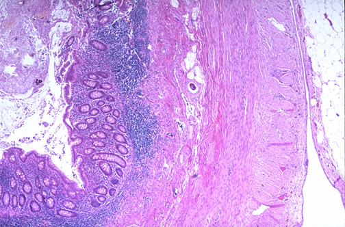
HISTOPATHOLOGY Histopathology is a study related to microscopic examination of a
LABORATORY biopsy or surgical specimen that has been processed and fixed onto
glass slides in order to analyze the signs of disease.
Using pathobiology understanding to evaluate and interpret the
shapes, sizes, and results of the cells and tissues architecture patterns
within a particular clinical context to get an accurate diagnosis.
Histological Techniques
Histological techniques are a Techniques that should be 1) Formalin-Fixed Paraffin-
series of crucial and important mastered: Embedded
Once the sections are prepared, they
procedures to produce Tissue grossing & fixation are usually stained to help distinguish
stained tissue samples on Tissue processing the components of the tissue.
glass slides for microscopic Tissue embedding 2) Frozen Section
Tissue sectioning Tissues are frozen rapidly, and then
examination. Tissue staining cut in a cryostat with a cold knife,
Slide mounting and labelling then stained and observed under the
microscope.
Equipment in Histopathology Laboratory
TISSUE PROCESSOR
Tissue Rotating MICROTOME
transfer head
basket Arm
attachment Adjustments Block
holder
slot Wax Stage Blade
Reagent container holder
containers Base
Keypad
control
EMBEDDING CENTRE
Wax reservoir Mould bin
Wax dispenser Forceps warmer
Cold plate Hot plate Tissue
warmer
ROUTINE WORKFLOW
Specimen fixed in 10% formalin tissue grossing tissue processing
Fixation status of a Specimen is grossed into 2-3mm Total tissue processing time
specimen is checked. sizes and kept in a cassette. takes about 21 hours.
floating out tissue section tissue sectioning tissue embedding
Tissue section is floated out in Tissue block is cut first, frozen and Tissue embedded in a block
water bath at 43°C-45°C. sectioned into thin ribbon of tissues. of paraffin wax.
fishing out tissue slide drying dewaxing using hot plate at 60°C
The best tissue section is selected Slides were allowed to Slides were dewaxed for
and separated by using an air-dried on the slide rack. approximately 4 minutes when
applicator stick.
drying slide using a hot plate.
SLIDe mounting and labelling
tissue staining
Stained-tissue slides were Stained-slides were allowed Using hematoxylin and eosin
mounted and labelled completely. to dry before mounting. (H&E) stain.
Farah Adriana
TISSUE PREPARATION Binti Mokhtar
pPrRinIcNipCleIPLE
Tissue samples are immersed in an adequate volume of fixative solution (10% neutral buffered formalin) to preserve them
from decaying or damage. The formalin has a property of forming cross links between proteins, thus stabilizing the tissue
structures. The neutral pH inhibits formation of formalin pigment. 1:10 to 1:20 volume ratio of tissue to fixative is necessary
for optimal fixation.
Grossing includes general inspection of tissue specimen and identification of normal and abnormal components. For
example, specimen from category C such as appendix requires simple dissection with sampling needing a low level of
diagnostic assessment/ preparation.
materials procedure
Human tissue specimens (liver, By using a pencil, cassettes were labelled clearly with the
tonsil, appendix, uterus) student's name/ ID number, type of specimen and date.
10% neutral buffered formalin
(commercially prepared)
Fixation status of the specimen was checked. Margin and
equipment
orientation of the specimen were also identified.
Ducted fume
hood.
Brand: IRYAS Relevant portions of the tissue specimen were selected
before grossing.
apparatus result
Scalpel holder Grossed By using a scalpel, specimen grossing was performed one
Scalpel blade tissue at a time.
Forceps
Dissecting board specimen
Tissue cassette Small tissue specimen (2-3 mm thick) was placed securely
Containers Tissue inside a plastic cassette.
MediSheet specimen
Kimwipes in cassette
Gloves Tissue cassette was then transferred into a container
Masks containing 10% Neutral Buffered Formalin (NBF).
problem and solution
All the remaining specimens were transferred back into
Uneven size of the specimen the specimen container for future references.
Choose a proper orientation of the Tissue grossing and fixation were performed in
the fume hood (grossing station) with appropriate
specimen before cutting.
PPE.
Improve technique of handling the Small tissue samples are usually fixed at room
scalpel.
Ensure that the thickness of a specimen temperature.
is no more than 3mm.
Time of fixation was at least 6 hours but not more
than 24-72 hours. This includes time in a processor
and documentation time.
Larger specimens may require longer time as
formalin slowly penetrates the tissues.
conclusion
Correct technique of tissue preparation (labelling cassette, grossing,
fixation) is very important to ensure good performance of subsequent
histology procedures such as embedding and sectioning.
Muhammad Syaddad
TISSUE PROCESSING Bin Zainuar
PRINCIPLE EQUIPMENT
Tissue processing is designed to remove all extractable water 3 Stages:
from the tissue, replacing it with a support medium that -Dehydration
provides sufficient rigidity to enable sectioning of the tissue -Clearing
without parenchymal damage or distortion. -Infiltration
APPARATUS MATERIALS Automated Tissue Processor
Brand: SHANDON CITADEL 1000
Forceps Tonsil, appendix and
Tissue processor organiser baskets uterus tissue specimen Model: SHA 69800001
Tissue processor basket lid fixed in 10% formalin.
Tissue processor reagent containers CHEMICAL/REAGENT
Measuring cylinder (1L & 2L)
Beaker (1L & 2L)
Gloves
Aprons
Masks
Goggles
PROCEDURE
Tissue blocks in cassettes that were fixed in 10% buffered formalin
were transferred and stacked onto tissue processor organiser baskets
by using forceps.
Tissue processor baskets filled with cassettes then were inserted into RESULT
the basket hanger with tissue processor basket lid positioned on top
Tissue block in
of it. cassette after
The tissue processor operating head door were open for the complete cycle of
attachment of the basket hangers by securing it into the operating processing.
head slot. PROBLEM
The processing time was confirmed as programmed as on hand-held
controller display & “AUTO START” key were pressed to commenced After a few cycles of tissue processing, the
reagent in the container starts to decrease in
the tissue processing cycle. volume due to evaporation & turns murky as
the tissue block is being socked in the
Dehydration Clearing Infiltration reagents.
Step 2-7 Step 8-10 Step 11 & 12 SOLUTION/TROUBLESHOOTING
Water & aqueous Dehydrating The clearing agent
fixatives are extracted compounds are is entirely removed The chemical reagents were replaced after
from fixed tissues in removed from from the tissue and the fourth cycle with freshly prepared
this procedure. the tissue and substituted with a reagents to ensure the next cycle of
To avoid distortion to replaced with a medium (wax) that specimen blocks were fully covered and
fragile tissue, wax-soluble completely fills all optimized the tissue processing process.
specimens are treated solution. of the tissue voids.
using a graded system The tissue then It provides a
of reagents with have translucent matrix and avoids
increasing appearance. tissue structural
concentrations. damage during
microtomy.
CONCLUSION
Although tissue processing is a very time consuming procedure, it is a very crucial step to emphasize as it
would determine the quality of tissue block specimens whether it is good enough to be carried for the
next steps (embedding, sectioning & staining) without any further problems.
Hajar Hanani
TISSUE EMBEDDING Binti Hishamudin
EQUIPMENT MATERIALS CHEMICAL/REAGENT
Paraffin Wax Tissue Embedding Centre Processed tissue blocks Paraffin wax
Brand : Slee Mainz MPS/C/MPS/P/MPS/W (liver, tonsil, appendix, uterus) Brand: SAKURA Tissue-Tek
Paraffin Wax
principle
APPARATUS Embedding is a process which will hold
the processed tissue in place by
Forceps Steel block Plastic cassette Gloves embedding the tissue into a block of
Scraper mould Tissue paper Masks paraffin wax. The tissue specimen was
enclosed in an embedding medium
Processed tissue procedure Tissue sample in using a mould. The embedding medium
cassette was placed the cassette was that being used is paraffin wax. The
Cryo console being viewed first medium will be able to support the
in the and the light embedded tissue blocks during
cassette bath were turned on The best mould microtomy in order to get a thin ribbon
that corresponds of tissue sections.
Processed tissue then A small amount of to the tissue’s size
transferred from the molten paraffin results
was poured into was chosen
cassette into the the mould Paraffin-
mould by using warm embedded
forceps tissue
blocks
The mould was The tissue was pressed More molten wax
transferred to gently and firmed into the was poured into problems and solution/troubleshooting
the cold plate wax using a pair of warm the mould until
forceps until the paraffin Wrong orientation of the processed tissue
almost full
solidified in a thin layer Check for the correct orientation of the
tissue beforehand proceed transferred
The mould was Molten wax was Labelled cassette and into the paraffin wax-mould
immediately being poured into the paper were placed on
cooled down by mould until it fully unstable cassette
placing it on the cold covered the face top of the mould.
of the cassette Re-embedded the
plate tissue in the
paraffin wax by
The paraffin tissue block was separated from dewax the
its mould after the paraffin wax solidified previous wax on
the hot surface of
Excess wax from paraffin tissue block cassette was embedding centre
removed by using scalpel.
Air bubbles are entrapped around
conclusion the tissue
Embedding step need to be done very carefully as the blocks' result can Embedded the tissue properly in the
also affecting tissue sectioning process. So, during embedding the molten wax by slowly pouring the
tissue specimen, sturdy hands needed and filled up the molten paraffin molten wax into the mould.
wax slowly to prevent from creating any bubbles, plus placing the
cassette correctly.
Sabrina Valeriana
TISSUE SECTIONING Senai Binti MacDonald
prpirnincicpipllee equipment
Rotary microtomes play an important role as they are capable of cutting sections Rotary microtome Slee Mainz CUT 5062
from paraffin blocks as thin as 1μm. As the block goes through the blade, a
segment with such a thickness will be created. When the sections are placed in a
water bath, the wax expands due to surface tension and heat, which aids in the
removal of creases and folds. Sharp and blemish-free blades are required for
satisfactory cutting. A good blade may section poorly prepared paraffin blocks,
but a bad blade can fail to cut even the best material.
apparatus material Floatation water bath XH-1001
Slide racks Paraffin block Hot plate or drying oven HPS-7C
Frosted slides (tonsil, appendix and uterus) Freezer
Brush
Gauze procedure
Pasteur pipettes
Applicator sticks 1.Setting up microtome 2. Trimming of tissue block
Pencil Blade angle was adjusted to Paraffin block was placed vertically or horizontally onto the cassette
Forceps 90 degree. Microtome bladder holder. Pressed forward the paraffin block until it touches the edge of the
Thermometer was placed which is a different knife.
site for trimming and Section thickness was set for 10-20 microns for large tissue pieces; 5-10
result sectioning. microns for small tissue pieces.
Blade fixations was tightened Stopped trimming once complete tissue sections appeared on the
by the right lever clockwise. section ribbon. Trimmed block was replaced in the freezer at -20 degree
and moved to an unused area bladder or placed a new bladder.
Ribbon section
Floating out section 4. Floating out sections 3. Cutting sections
Ribbon sections was gently pressed Took out the paraffin block from the freezer. Placed paraffin
by using an applicator stick to block onto the cassette holder.
prevent wrinkles or bubbles. The Changed the “Mode” and thickness of 5-10 microns was
best section from the ribbon was selected.
selected and separated it by using A series of paraffin sections was cut until produce a ribbon of
an applicator stick. serial sections. By using a brush, ribbon section from the knife
Make sure not to leave sections of edge was separated. Ribbon sections was transferred into
ribbon too long to prevent tissues flotation water bath that filled with distilled water (42°C-45°C)
expanding and distorted.
5. Picking up sections 6. Drying sections
Sections was collected using a clean slide by using The slides were allowed to dry on the
fishing techniques. The slide was immersed into the slides rack at room temperature. Any
water bath to get the best result. water was removed from the slides
The slide was held vertically beneath the section and before proceeding to the drying oven.
lifted up gently to make sure tissue adherence to the
slide.
Tissuperisneccitpiolneingcios naclturuseioanrt that requires a problem & solution/troubleshoot
great deal of talent and experience. Good Tissue ribbons are not form
sectioning will give the best result in visualizing Paraffin block may be unstable.
tissue under the microscope. However, it is
important to have properly fixed and embedded Cooling paraffin block to stabilpizreinit.ciple
blocks or artefacts including tearing, ripping,
holes or folding will appear. Large holes formed in sections
Tissue is not embedded properly
Depending on issue/problems, Re-embed
tissue as required.
Muhamad Zul Azmeer
TISSUE STAINING Bin Mohd Nasir
PRINCIPLE
In general, tissue staining is used to emphasize the tissue components and improve tissue contrast. In histological studies,
the H&E staining is used to distinguish cell components, as H&E dyes contrasting colour for cell’s nucleus and cytoplasm
components.
Hematoxylin is a basic dye, which stains the cell’s nucleus, gives it bluish colour.
Eosin is an acidic dye, which stains the cytoplasm components of cell, gives it a pink-reddish colour.
CHEMICALS/REAGENTS APPARATUS EQUIPMENTS
Hematoxylin 3G (Sakura) - Commercially Slide racks Oven
prepared Reagent containers Brand: Memmert
Eosin (Sakura) - Commercially prepared Forceps Hotplate & Stirrer
Xylene Filter paper
Alcohol (Absolute, 95%) Tissue paper Brand: Pro
Distilled water Funnel Model: HP-7 Lab
Tap water Measuring cylinder
Tripod stand Plus Series
MATERIALS
Unstained tissue slides
PROCEDURE
Deparaffinization Hydration . Nucleus staining
The tissue section was dewaxed by The tissue section was passed through: Tissue section was stained
using:
2 changes of absolute alcohol, 1 with hematoxylin 3G
The oven for 30 minute each station. (Sakura), for 5 minutes.
minutes/hotplate for 4 minutes A change of 95% alcohol, for 1
each slides at 60℃. minute Bluing/Washing
3 changes of xylene, 3 minutes Running tap water, for 1 minutes The tissue section was passed
each station. Distilled water, for 1 minutes through:
Washing Cytoplasm staining Running tap water, for 5
The tissue section was passed through: The tissue section was stained minutes.
Distilled water, for 30
Distilled water, for 10 seconds. with eosin (Sakura), for 2 seconds for excess stain
2 changes of 95% Alcohol, for 10 minutes removal.
seconds each station.
Dehydration Clearing
The tissue section was passed The tissue section was passed
through: through:
3 changes of absolute alcohol, for 2 changes of xylene, 1 minute
1 minute each station and 2 minutes for each station
RESULTS PROBLEM CONCLUSION
Stained Irregular staining of the tissue section H&E staining, known as a standard
tissue due to the incompletely paraffin removal staining protocol of tissue staining.
slides It is one of the vital procedure in
Stained during deparaffinization. histological studies, commonly
tissue used to differentiate the features
observed SOLUTION/TROUBLESHOOTING in cell, nucleus and cytoplasmic
under components. Its dyes, hematoxylin
microscope Ensure to follow the time for xylene, OR will stain the nucleus and gives the
bluish colour. On the other hand,
Replace the xylene with the new one, as it eosin will stain the cytoplasmic
can increase the deparaffinized time. components and gives the pink-
reddish colour.
Farah Adriana Binti Mokhtar
SLIDE MOUNTING & LABELLING
PRINCIPLE
Tissue sections that need to be examined at any length of time or to be stored for a specific duration of time
must be mounted using appropriate mounting media. Mounting media are used to protect and preserve tissue
sections and to enhance the microscopic evaluation of the tissue. A mounting medium with an refractive index
close to that of the fixed tissue will provide a transparency, with only the stained tissue elements visible. The
mounting medium used in the laboratory will harden to hold the coverslip firmly in place.
MATERIALS CHEMICAL/REAGENT EQUIPMENT
H&E stained slides Mounting medium Ductless fume hood
Product's name: CoverSeal-X Brand: ESCO
APPARATUS Angle for slide mounting Model: Ascent Max
Slide holders Image retrieved from Slide Player in Image retrieved from
Coverslips (22 x 40 mm / 24 x 60 mm) Microscopy Cell Structure Mitosis Youtube MedLab
Applicator sticks
MediSheets Channel, Mounting &
Self-adhesive label stickers Labelling
PROCEDURE result
A self-adhesive sticker was labelled with the following Mounted H&E
details: Student’s name, Student’s ID number, Type of stained slide
with a label
specimen, Type of stain and Date.
PROBLEM & SOLUTON
A coverslip was chosen appropriately according to the Air bubbles entrapped under the
size of the tissue section. coverslip
Gently press the coverslip by using an
A mounting medium was placed adequately on one applicator stick to remove the air bubble and
edge of the coverslip. evenly spread the mounting medium under
the coverslip.
Coverslip was holded at 45 degree and was gently Excess mounting medium
lowered until it was placed on the slide. Capillary flowing out of the slide.
attraction will cause the mounting medium to flow
Let the mounted slide dry and
upwards, carrying the coverslip along with it. then use a scalpel to remove
the excess mounting medium.
The slide was examined microscopically to make sure CONCLUSION
there were no air bubbles.
Mounting should be done properly with
less error concerning air bubbles.
The coverslip was pressed gently using an applicator stick Complete labelling is a must to provide
to remove the air bubbles. reliable result when evaluating the
tissue section.
The written sticker label was pasted at the frosted
end of the slide.
Finally, the slide was placed on the slide holder to be air-
dried.
Zul Azmeer, Syaddad, Hajar Hanani, Valeriana
FROZEN SECTION
principle CHEMICAL/REAGENT
The tissue specimen is rapidly frozen by cryostat which converting water to Tissue Freezing Medium /
ice, that serves as an embedding medium and allows the tissue to be Frozen Section Compound
10% Neutral Buffered Formalin
sectioned (cryotomy). The temperature of the tissue sample is decreased to Hematoxylin 3G (Sakura)
-20°C, resulting to a stiffer tissue sample. Eosin (Sakura)
Alcohol (95% & Absolute)
EQUIPMENT APPARATUS Xylene
Tap water
Cryostat (brand: Slee Mainz MEV) Glass slide Distilled water
Oven (brand: Memmert) Coverslip
Microtome MATERIALS
Blade
Forceps Fresh tissue specimen
Brush
Applicator stick
Coplin jar
Gloves
procedure results
Two slides were labelled with the surgical pathology "FS" and the medical
record number.
A cryo cassette was constructed. A tiny amount of O.C.T. cryomatrix should Ribbon sectioned Stained tissue slide
be added. The cryocassette was placed on the cryobar and the cryospray was
used on it to freeze. Adapted from Stephen R. Peters M.D. (2015) in Frozen
Section Techniques
The block was trimmed carefully at 15 micron intervals until the entire surface of
the tissue is revealed. The knife was wiped gently by using a brush. Then, a problems and
portion was cut about 4-5 microns thick.. solutions/troubleshooting
The section was extracted from the knife by using a coated glass slide at Ice crystal artifacts
room temperature. Then, two (2) frozen section slides each block were
stained with H&E and immediately transferred to hematoxylin. Increase the freezing time of the
tissue, as it can reduce the size
Stained in hematoxylin for 1 minute. Rinsed in tap water. of ice crystal artifacts. Therefore,
1 % Acid alcohol 1-2 dipped. Rinsed in tap water the tissue damage can be
Stained in 1% Alcoholic Eosin for 30 seconds. Rinsed in tap water minimized.
Dehydrated in 3 different Alcohol
Cleared in 3 changes of Xylene Tissue tears or streaks
Mounted with DPX mountant and coverslip.
Check the entire blade as it may
conclusion due to the presence of frozen
debris stuck on it, OR
Frozen section is an alternative method of paraffin section in tissue
diagnosis. It is very useful in dealing with STAT (Short Turn Around Replace the blade with the new
Time) specimen as it consists of rapid procedures compared to the one.
paraffin section. However, it has a strict regulation likes a
temperature sensitivity of the tissue sample. Therefore, it is needed
to follow each step of the procedures to reduce a possibility of
tissue damage.
REFERENCES
Ahmed, Mahtab. (2016). Steps of tissue processing in histopathology laboratory, Review Report. HEALTH DIGEST. 1. 26-27.
Al-Sabaawy, H. B. (2021, December 1). Standard techniques for formalin-fixed paraffin-embedded tissue: A Pathologist’s
perspective. Iraqi Journal of Veterinary Sciences. Retrieved from https://vetmedmosul.com/article_169996.html
Alturkistani, H. A., Tashkandi, F. M., & Mohammedsaleh, Z. M. (2016). Histological stains: a literature review and case study.
Global journal of health science, 8(3), 72. Retrieved from https://www.ncbi.nlm.nih.gov/pmc/articles/PMC4804027/
Gurcan, M. N., Boucheron, L. E., Can, A., Madabhushi, A., Rajpoot, N. M., & Yener, B. "Histopathological Image Analysis: A
Review," in IEEE Reviews in Biomedical Engineering, vol. 2, pp. 147-171, 2009, doi: 10.1109/RBME.2009.2034865.
Jose, M., & Adyanthaya, S. (2013). Quality and safety aspects in histopathology laboratory. Journal of Oral and Maxillofacial
Pathology, 17(3), 402–407. https://doi.org/10.4103/0973-029x.125207
National Society For Histotechnology 1973. (2001, July). Guidelines for Hematoxylin and Eosin Staining. Retrieved from
http://nsh.org/sites/default/files/Guidelines_For_Hematoxylin_and_Eosin_Staining.pdf
Paxton, S. (2003). What is Histology: The Histology Guide. The Histology Guide, University of Leeds. Retrieved from
http://histology.leeds.ac.uk/what-is-histology/histological_sections.php
Ravikumar, S., Surekha, R., & Thavarajah, R. (2014). Mounting media: An overview. Journal of Dr. NTR University of Health
Sciences, 3(5), 1. https://doi.org/10.4103/2277-8632.128479
Rolls, G. (2016, June 16). Steps to Better Grossing. Leica Biosystems; Leica Biosystems. Retrieved from
https://www.leicabiosystems.com/knowledge-pathway/steps-to-better-grossing/
Rolls, G., & Sampias, C. (n.d.). H&E Staining Overview: A Guide to Best Practices. Leica Biosystems. Retrieved from
https://www.leicabiosystems.com/knowledge-pathway/he-staining-overview-a-guide-to-best-practices/
Rolls, G. B. (2019, April 15). An Introduction to Specimen Processing. Leica Biosystems. Retrieved from
https://www.leicabiosystems.com/knowledge-pathway/an-introduction-to-specimen-processing/
Russell, K. (2014). Microscopy Cell Structure Mitosis. Slide Player. https://slideplayer.com/slide/276707/
S. (2014, May 4). Embedding. MODULE Histology and Cytology. Retrieved from
https://nios.ac.in/media/documents/dmlt/HC/Lesson-08.pdf
Sim, J. (2019, November 13). What is a Frozen Section? Biorepository. Retrieved from https://www.geneticistinc.com/blog/what-is-
a-frozen-section
Slaoui, Mohamed & Fiette, Laurence. (2011). Histopathology Procedures: From Tissue Sampling to Histopathological Evaluation.
Methods in molecular biology (Clifton, N.J.). 691. 69-82. 10.1007/978-1-60761-849-2_4.
Themes, U. (2017, December 13). Tissue processing. Basicmedical Key. Retrieved from https://basicmedicalkey.com/tissue-
processing/
Wallace, J. (2011, October 1). Frozen section procedure. PathologyOutlines.com. Retrieved from
https://www.pathologyoutlines.com/topic/breastfrozenprocedure.html.
Special Appreciation To
Dr Nur Ayunie Binti Zulkepli
Madam Hartini Yusof
Sir Mohd Nornizam Bin Ahmad Zaini
Miss Salina Binti Shafie
Sir Mohd Nazzihan Md Ajis
The words you are searching are inside this book. To get more targeted content, please make full-text search by clicking here.
1.MUHAMAD ZUL AZMEER BIN MOHD NASIR (2020473736)
2.MUHAMMAD SYADDAD BIN ZAINUAR (2020846902)
3.FARAH ADRIANA BINTI MOKHTAR (2020473348)
4.HAJAR HANANI BINTI HISHAMUDIN (2020492628)
5.SABRINA VALERIANA SENAI BINTI MACDONALD (2020828002)
Discover the best professional documents and content resources in AnyFlip Document Base.
Search
HS2413B_HISTOLOGICAL LAB REPORT GROUP 2
- 1 - 11
Pages: